Fovea centralis
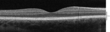
[4] The fovea is a depression in the inner retinal surface, about 1.5 mm wide, the photoreceptor layer of which is entirely cones and which is specialized for maximum visual acuity.
[5] The fovea is located in a small avascular zone and receives most of its oxygen from the vessels in the choroid, which is across the retinal pigment epithelium and Bruch's membrane.
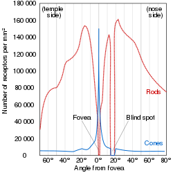
[6] The center of the fovea is the foveola – about 0.35 mm in diameter – or central pit where only cone photoreceptors are present and there are virtually no rods.
Starting at the outskirts of the fovea, however, rods gradually appear, and the absolute density of cone receptors progressively decreases.
In 2018, the anatomy of the foveola was reinvestigated, and it was discovered that outer segments from the central foveolar cones of monkeys are not straight and twice as long as those from the parafovea.
[11] Therefore, the acuity of foveal vision is limited only by the density of the cone mosaic, and the fovea is the area of the eye with the highest sensitivity to fine details.
[13] The fovea sees only the central two degrees of the visual field, (approximately twice the width of your thumbnail at arm's length).
Foveal fixation is also considered as a overt form of attention which allows to focus sensory processing resources on the most relevant sources of information.
They are concentrated in the Henle fiber layer (photoreceptor axons that go radially outward from the fovea) and to a lesser extent in the cones.
[23][24] They are believed to play a protective role against the effects of high intensities of blue light which can damage the sensitive cones.
[28] From these values, one can calculate the average angle of view of a single sensor (cone cell), which is approximately 31.46 arc seconds.
[29] Assuming average focal lengths, this suggests that individuals with both high cone densities and perfect optics may resolve pixels with an angular size of 21.2 arc seconds, requiring PPI values at least 1.5 times those shown above in order for images not to appear pixelated.
The presence of the pigment in the radially arranged axons of the Henle fiber layer causes it to be dichroic and birefringent[30] to blue light.
Among mammals, it is found in its most developed form only in Haplorhine primates, although a more rudimentary fovea-like structure exists in some diurnal lemurs.