Leukoaraiosis
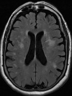
[1][2] On MRI, leukoaraiosis changes appear as white matter hyperintensities (WMHs) in T2 FLAIR images.
[3][4] On CT scans, leukoaraiosis appears as hypodense periventricular white-matter lesions.
[5] The term "leukoaraiosis" was coined in 1986[6][7] by Hachinski, Potter, and Merskey as a descriptive term for rarefaction ("araiosis") of the white matter, showing up as decreased density on CT and increased signal intensity on T2/FLAIR sequences (white matter hyperintensities) performed as part of MRI brain scans.
Hypertension, smoking, diabetes,[3] hyperhomocysteinemia, and heart diseases are all risk factors for leukoaraiosis.
White matter hyperintensities can be caused by a variety of factors, including ischemia, micro-hemorrhages, gliosis, damage to small blood vessel walls, breaches of the barrier between the cerebrospinal fluid and the brain, or loss and deformation of the myelin sheath.