Magnetosome
Each magnetosome can often contain 15 to 20 magnetite crystals that form a chain which acts like a compass needle to orient magnetotactic bacteria in geomagnetic fields, thereby simplifying their search for their preferred microaerophilic environments.
[2] Magnetite-bearing magnetosomes have also been found in eukaryotic magnetotactic algae, with each cell containing several thousand crystals.
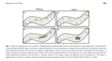
[3][4] Magnetotactic bacteria are widespread, motile, diverse prokaryotes that biomineralize a unique organelle called the magnetosome.
A magnetosome consists of a nano-sized crystal of a magnetic iron mineral, which is enveloped by a lipid bilayer membrane.
[6] Biosynthesis of magnetite particles in vertebrates like mammals is implied to be similar to that observed in bacterial cells, although no evidence is provided.
Finally, the human magnetosomic organelle has an unknown function that does not involve detecting the earth's magnetic field.
[citation needed] Magnetotactic bacteria use a process known as biomineralization to exert an incredible degree of control on the formation of the mineral crystals within the magnetosomes.
[10] The arrangement of the magnetites is critical because individually they are not very strong, but when linked in an ordered chain they increase significantly in strength.
Certain subgroups of the Pseudomonadota in the domain of Bacteria have been found through analyses of the MTB’s RNA to only use iron oxide which is the more common material.
These non-ideal arrangements may lead to additional, currently unknown functions of magnetosomes; possibly related to metabolism.
The magnetosome shape and elastic properties of biological membranes are what is holding the chains together, as well as the linearity and the connection to the cytoskeleton.
The cell wall and associated membrane structures have been thought to act to prevent magnetosome chain collapse.
![{\displaystyle \scriptstyle [1{\overline {1}}0]}](https://wikimedia.org/api/rest_v1/media/math/render/svg/f99ce4c507bedc2d8c9e600715fe15e2d2666907)