Male reproductive system
The main male sex organs are the penis and the scrotum, which contains the testicles that produce semen and sperm, which, as part of sexual intercourse, fertilize an ovum in the female's body; the fertilized ovum (zygote) develops into a fetus, which is later born as an infant.
The penis is an intromittent organ with a long shaft, an enlarged bulbous-shaped tip called the glans and its foreskin for protection.
When a male becomes sexually aroused, erection occurs because sinuses within the erectile tissues of the penis (corpora cavernosa and corpus spongiosum) become filled with blood.
The arteries of the penis are dilated while the veins are compressed so that blood flows into the erectile cartilage under pressure.
During times of lower temperatures, the cremaster muscle contracts and pulls the scrotum closer to the body, while the dartos fascia gives it a wrinkled appearance; when the temperature increases, the cremaster and dartos fascia relax to bring down the scrotum away from the body and remove the wrinkles respectively.
The testicles are two gonads that produce sperm by meiotic division of germ cells within the seminiferous tubules,[1] and synthesize and secrete androgens that regulate the male reproductive functions.
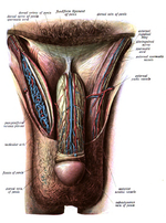
[2] The vas deferens, which is also known as the sperm duct, is a thin tube approximately 30 centimetres (0.98 ft) long that starts from the epididymis to the pelvic cavity.
Three accessory glands provide fluids that lubricate the duct system and nourish the sperm cells.
It begins with a single fertilized egg and culminates 38 weeks later with the birth of a male child.
Sexual identity is determined at fertilization when the genetic sex of the zygote has been initialized by a sperm cell containing either an X or Y chromosome.
A sperm cell carrying a Y chromosome results in an XY combination, and a male child will develop.
Once inside the target cells, testosterone is converted by means of an enzyme called 5α-reductase into the dihydrotestosterone (DHT).
Internal changes include the formation of the tubular seminar Chris tubules in the rete testis from the primary sex cord.
Descent does not occur until about the 28th week when compared to when canals form and the abdominal wall provides openings from the pelvic cavity to the scrotal sac.
The urethral groove forms on the ventral surface of the phallus early in development during the differentiation of the external genitalia.
This sequence is understandable in light of the fact that both male and female embryos develop within the maternal environment - high in estrogen secreted by the mother's ovaries and the placenta.
[10] During puberty, increased gonadotropin secretion stimulates a rise in sex steroids creation from the testes.
The increased secretion of testosterone from the testes during puberty causes the male secondary sexual characteristics to be manifested.
The gene for sexual differentiation in humans, called the testis determining factor (TDF),[21][non-primary source needed] is located on the short arm of the Y chromosome.
By week six, string-like cell congregations called primitive sex cords form within the enlarging genital ridge.
[4] Specialized primordial germ cells are forming and migrating from the yolk sac to the embryonic gonads during week eight and nine.
Testosterone secretion by the interstitial cells of the testes then causes the growth and development of the mesonephric ducts into male secondary sex organs.
[7] The Müllerian ducts atrophy, but traces of their anterior ends are represented by the appendices testis (hydatids of Morgagni of the male), while their terminal fused portions form the utriculus on the floor of the prostatic urethra.


* w, w. Right and left Wolffian ducts.

