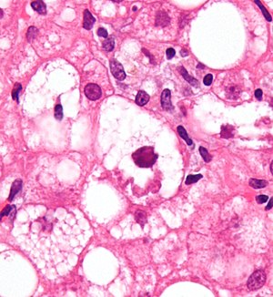
Mallory body
[1] Mallory bodies are damaged intermediate filaments within the liver cells.
[3] They are a recognized feature of Wilson's disease (25%), primary biliary cirrhosis (24%), non-alcoholic cirrhosis (24%), hepatocellular carcinoma (23%) and morbid obesity (8%), among other conditions.
[4] Mallory bodies are highly eosinophilic and thus appear pink on H&E stain.
[5] It is named for the American pathologist Frank Burr Mallory, who first described the structures in 1911.
[3] A renaming as Mallory–Denk bodies was proposed in 2007 to honor the contribution of Austrian pathologist Helmut Denk for the molecular analysis of the pathogenesis of MDBs.