Melanocyte
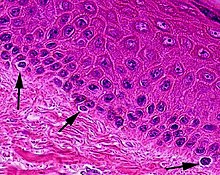
Melanocytes are melanin-producing neural crest-derived[3] cells located in the bottom layer (the stratum basale) of the skin's epidermis, the middle layer of the eye (the uvea),[4] the inner ear,[5] vaginal epithelium,[6] meninges,[7] bones,[8] and heart found in many mammals and birds.
Once synthesized, melanin is contained in special organelles called melanosomes which can be transported to nearby keratinocytes to induce pigmentation.
Through a process called melanogenesis, melanocytes produce melanin, which is a pigment found in the skin, eyes, hair, nasal cavity, and inner ear.
The color of the melanin is black, allowing it to absorb a majority of the UV light and block it from passing through the epidermis.
Typically, between 1000 and 2000 melanocytes are found per square millimeter of skin or approximately 5% to 10% of the cells in the basal layer of epidermis.
Importantly, melanocytes stimulated by cytokines express surface proteins such as CD40 and ICAM1 in addition to MHC class II, allowing for co-stimulation of T cells.
[21] They likely work in concert with both keratinocytes and Langerhans cells,[20][21] both of which are also actively phagocytic,[23] to contribute to the immune response.
The copper-ion based enzyme-catalyzed oxidative transformation of catechol derivative dopa to light absorbing dopaquinone to indole-5,6-quinone is clearly seen following the polymerization to melanin, the color of the pigment ranges from red to dark brown.
Numerous stimuli are able to alter melanogenesis, or the production of melanin by cultured melanocytes, although the method by which it works is not fully understood.
Increased melanin production is seen in conditions where adrenocorticotropic hormone (ACTH) is elevated, such as Addison's and Cushing's disease.
When ultraviolet rays penetrate the skin and damage DNA, thymidine dinucleotide (pTpT) fragments from damaged DNA will trigger melanogenesis[26] and cause the melanocyte to produce melanosomes, which are then transferred by dendrites to the top layer of keratinocytes.
In adults, stem cells are contained in the bulge area of the outer root sheath of hair follicles.