Melioidosis
The disease should be considered in anyone who has spent time in endemic areas who develops a fever, pneumonia, or abscesses in their liver, spleen, prostate, or parotid gland.
[2] Results of a chest X-ray can range from diffuse nodular infiltrates in those with septic shock to progressive consolidation located most commonly in the upper lobes for those with pneumonia only.
[1] Rare manifestations include lymph node disease resembling tuberculosis,[10] mediastinal masses, pericardial effusion,[3] mycotic aneurysm,[1] and inflammation of the pancreas.
Those with melioidosis encephalomyelitis tend to have normal computed tomography (CT) scans but increased T2 signal by magnetic resonance imaging (MRI), extending to the brain stem and spinal cord.
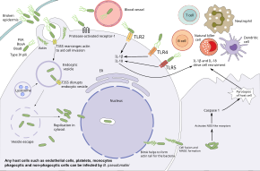
[10] Additionally, adhesion of the bacteria partially depends on the presence of the host protein Protease-activated receptor-1 which is present on the surface of endothelial cells, platelets, and monocytes.
When MNGCs lyse, they form plaques (a central clear area with a ring of fused cells) that provide shelter for the bacteria for further replication or latent infection.
Macrophages activated by interferon gamma (IFN) have improved the killing of B. pseudomallei via the production of inducible nitric oxide synthase.
[1][10] Additional elements of the immune system are activated by the host toll-like receptors such as TLR2, TLR4, and TLR5 that recognize the conserved pieces of the bacteria such as LPS and flagella.
Although macrophages show deregulated cytokine responses in individuals with HIV infection, bacterial internalization and intracellular killing are still effective.
Amongst mechanisms suggested are: residing in the nucleus of the cell to prevent being digested, entering a stage of slower growth, antibiotic resistance, and genetic adaption to the host environment.
Other methods of antigen detection such as direct immunofluorescence, antibody-sandwich ELISAs, and lateral flow immunoassays using monoclonal antibody.
[32] In the United States, lab workers can handle clinical specimens of B. pseudomallei under BSL-2 conditions, while mass production of such organisms requires BSL-3 precautions.
[1] Those staying in endemic areas should avoid direct contact with soil and outdoor exposure to heavy rain or dust clouds.
[41] Before 1989, the standard treatment for acute melioidosis was a three-drug combination of chloramphenicol, co-trimoxazole, and doxycycline; this regimen is associated with a mortality rate of 80% and is no longer used unless no other alternatives are available.
Co-trimoxazole is recommended in addition to ceftazidime for neurological melioidosis, osteomyelitis, septic arthritis, skin and gastrointestinal infection, and deeply seated abscesses.
For deep-seated infections such as abscesses of internal organs, osteomyelitis, septic arthritis, and neurological melioidosis, the duration of antibiotics given should be longer (up to 4 to 8 weeks).
Co-amoxiclav is an alternative for patients unable to take co-trimoxazole and doxycycline (e.g. pregnant women and children under the age of 12), but is not as effective and has a higher relapse rate.
[1][46] There are also cases where melioidosis is successfully treated with co-trimoxazole for 3 months without going through intensive therapy provided that there is only skin manifestations without the involvement of internal organs or sepsis.
[54][55][56] Several immunomodulating therapies are suggested to boost the human body's immune function against the bacteria because the pathogenesis of melioidosis is thought to be contributed by defects in neutrophils.
[1] The Royal Darwin Hospital 2014 guidelines recommended granulocyte colony-stimulating factor (G-CSF) as immunomodulating therapy for those with septic shock at 300 ug daily as soon as the bacteriological laboratory flag the culture as possibly Burkholderia pseudomallei.
The G-CSF is continued for ten days depending on clinical response or a contraindication develops such as a white cell count greater than >50,000 X106/litre.
[59] Underlying medical conditions such as diabetes mellitus, chronic kidney disease, and cancer can worsen the long-term survival and disability of those who recover from infection.
[61][62] Melioidosis is endemic in parts of southeast Asia (including Thailand,[63] Laos,[64] Singapore,[65] Brunei,[66] Malaysia,[67] Myanmar[68] and Vietnam[69]), southern China,[70] Taiwan[71] northern Australia.
One possible explanation is that importation of medicinal plant products or exotic reptiles could have resulted in the introduction of melioidosis in the United States.
Excessive release of Tumor necrosis factor alpha and Interleukin 12 by mononuclear cells increases the risk of septic shock.
For example, 25% of children started producing antibodies against B. pseudomallei between 6 months to 4 years of staying in endemic areas although they did not experience any melioidosis symptoms; suggesting they were exposed to it over this time.
[15] In the same year, melioidosis outbreak occurred inside the Institute for Medical Research (IMR), Kuala Lumpur, Malaya after its laboratory animals such as guinea pigs and rabbits were infected.
It was only in 1917 when Fletcher isolated an organism similar to Whitmore's bacillus from a Tamil rubber estate worker, the presence of the new species of bacteria was confirmed.
[90] However, this hypothesis was later disproved in 2017 when whole genome sequencing of B. pseudomallei over 30 countries collected over 79 years suggested Australia as the early reservoir for melioidosis.
[94] An outbreak of melioidosis at the Paris Zoo in the 1970s (known as L'affaire du jardin des plantes) was thought to have originated from an imported panda or horses from Iran.