Tobacco mosaic virus
[2][3] In 1886, Adolf Mayer first described the tobacco mosaic disease that could be transferred between plants, similar to bacterial infections.
[7] However, Ivanovsky remained rather convinced, despite repeated failures to produce evidence, that the causal agent was an unculturable bacterium, too small to be retained on the employed Chamberland filters and to be detected in the light microscope.
In 1898, Martinus Beijerinck independently replicated Ivanovsky's filtration experiments and then showed that the infectious agent was able to reproduce and multiply in the host cells of the tobacco plant.
[5][8] Beijerinck adopted the term of "virus" to indicate that the causal agent of tobacco mosaic disease was of non-bacterial nature.
[5] For his work, he was awarded 1/4 of the Nobel Prize in Chemistry in 1946,[10][11] even though it was later shown some of his conclusions (in particular, that the crystals were pure protein, and assembled by autocatalysis) were incorrect.
[13] In 1955, Heinz Fraenkel-Conrat and Robley Williams showed that purified TMV RNA and its capsid (coat) protein assemble by themselves to functional viruses, indicating that this is the most stable structure (the one with the lowest free energy).
The crystallographer Rosalind Franklin worked for Stanley for about a month at Berkeley, and later designed and built a model of TMV for the 1958 World's Fair at Brussels.
[15] The investigations of tobacco mosaic disease and subsequent discovery of its viral nature were instrumental in the establishment of the general concepts of virology.
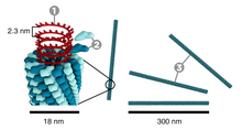
[16] The protein monomer consists of 158 amino acids which are assembled into four main alpha-helices, which are joined by a prominent loop proximal to the axis of the virion.
[18] X-ray fiber diffraction structure of the intact virus was studied based on an electron density map at 3.6 Å resolution.
This happens due to the formation of an obligatory intermediate produced from a protein allows the virus to recognize a specific RNA hairpin structure.
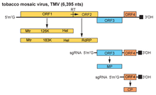
[24] The genome encodes 4 open reading frames (ORFs), two of which produce a single protein due to ribosomal readthrough of a leaky UAG stop codon.
Although TMV does not have defined transmission vectors, the virus can be easily transmitted from the infected hosts to the healthy plants by human handling.
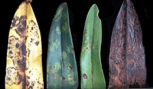
This is followed quickly by the development of a "mosaic" or mottled pattern of light and dark green areas in the leaves.
As long as the surrounding temperature remains below approximately 40 degrees Celsius, TMV can sustain its stable form.
If necessary, greenhouses and botanical gardens would provide the most favorable condition for TMV to spread out, due to the high population density of possible hosts and the constant temperature throughout the year.
Both tobacco and the beans P. vulgaris and V. sinensis suffered almost no lesioning in vitro from TMV when treated with a P. polycephalum extract.
Their research showed that salicylic acid most likely was disrupting replication and transcription and more specifically, the RdRp complex.
[42] The large amount of literature about TMV and its choice for many pioneering investigations in structural biology (including X-ray diffraction and X-ray crystallography), virus assembly and disassembly, and so on, are fundamentally due to the large quantities that can be obtained, plus the fact that it does not infect animals.
[31] James D. Watson, in his memoir The Double Helix, cites his x-ray investigation of TMV's helical structure as an important step in deducing the nature of the DNA molecule.
[45][46] Viral vectors based on TMV include those of the magnICON and TRBO plant expression technologies.
[48][49] The TMV-based vector also enabled C. acutatum to transiently express exogenous GFP up to six subcultures and for at least 2 mo after infection, without the need to develop transformation technology, RNAi can be expressed in the phytopathogenic fungus Colletotrichum acutatum by VIGS using a recombinant vector based on TMV in which the ORF of the gene encoding the green fluorescent protein (GFP) was transcribed in fungal cells from a duplicate of the TMV coat protein (CP) subgenomic mRNA promoter and demonstrated that the approach could be used to obtain foreign protein expression in fungi.