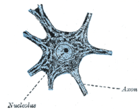
Nissl body
[2][3] The term "Nissl bodies" generally refers to discrete clumps of rough endoplasmic reticulum and free ribosomes in nerve cells.
Masses of rough endoplasmic reticulum also occur in some non-neuronal cells, where they are referred to as ergastoplasm, basophilic bodies,[1] or chromophilic substance.
[4] While these organelles differ in some ways from Nissl bodies in neurons,[5] large amounts of rough endoplasmic reticulum are generally linked to the copious production of proteins.
[1] "Nissl stains" refers to various basic dyes that selectively label negatively charged molecules such as DNA and RNA.
[6] They vary in size, shape, and intracellular location; they are most conspicuous in the motor neurons of the spinal cord and brainstem, where they appear as large, blocky assemblies.