Oligodendrocyte
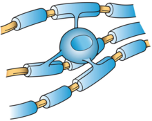
Oligodendrocytes (from Greek 'cells with a few branches'), also known as oligodendroglia, are a type of neuroglia whose main function is to provide the myelin sheath to neuronal axons in the central nervous system (CNS).
Studies have suggested that they originate from the ventral ventricular zone of the embryonic spinal cord, with some potential concentrations in the forebrain.
They arise during development from oligodendrocyte precursor cells (OPCs),[8] which can be identified by their expression of a number of antigens, including the ganglioside GD3,[9][10][11] the NG2 chondroitin sulfate proteoglycan, and the platelet-derived growth factor-alpha receptor subunit (PDGF-alphaR).
[20] Remarkably, oligodendrocyte population originated in the subventricular zone can be dramatically expanded by administering epidermal growth factor (EGF).
[21][22] Mammalian nervous systems depend crucially on myelin sheaths, which reduce ion leakage and decrease the capacitance of the cell membrane, for rapid signal conduction.
[23] Myelin also increases impulse speed, as saltatory conduction of action potentials occurs at the nodes of Ranvier in oligodendrocytes.
[24] Myelination is an important component of intelligence, and white matter quantity may be positively correlated with IQ test results in children.
[25] Oligodendrocytes, best known for their role in myelinating axons in the central nervous system, also have important functions in immune regulation.
The immature oligodendrocytes, which increase in number during mid-gestation, are more vulnerable to hypoxic injury and are involved in periventricular leukomalacia.
In cerebral palsy, spinal cord injury, stroke and possibly multiple sclerosis, oligodendrocytes are thought to be damaged by excessive release of the neurotransmitter, glutamate.
[36] Oligodendrocytes are also susceptible to infection by the JC virus, which causes progressive multifocal leukoencephalopathy (PML), a condition that specifically affects white matter, typically in immunocompromised patients.
[40] Oligodendrocytes also abundantly express components of the amyloidogenic pathway,[41][42][43] produce amyloid beta (Aβ), and contribute to plaque burden,[42][43] which is relevant when considering therapeutic interventions for Alzheimer's disease.