Opalinidae
[2] Most opalines live in the large intestine and cloaca of anurans (frogs and toads), though they are sometimes found in fish, reptiles, molluscs and insects; whether they are parasitic is not certain.
The unusual features of the opalines, first observed by Antonie van Leeuwenhoek in 1683,[3] has led to much debate regarding their phylogenetic position among the protists.
The relationship between opalines and other protists has been a subject of great controversy since the late 19th century, and is not completely resolved at present.
Initially, microscopists believed that the thousands of rhythmically beating hair-like structures which cover their surface were cilia, and they placed the opalines in Ciliophora.
[5] In the 1980s, detailed ultrastructural studies of Opalina ranarum revealed that they share many features with the heterokonts of the family Proteromonadidae.
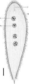
Two additional genera, Hegneriella Earl 1971 and Bezzenbergeria Earl 1973, have not been considered as valid by subsequent authors (p. 249)[2] The 5 recognized genera differ in terms of the number of nuclei, the appearance and location of the falx (two short, sickle-shaped rows of flagella), and whether the long rows of flagella (called "kineties") cover the body evenly or if there is a "bald spot".
[8] A more recent study found that Cepedea couillardi fits the standard opaline life cycle model described below, while that of Opalina proteus is completed entirely in the tadpole stage of the host.
At some point the small tomonts undergo encystment, and the cysts are released into the environment (i.e. the breeding pool of the anuran host) along with the feces.
One study found no irritation or other pathological signs on the rectal epithelium of Symphysodon aequifasciata infested with Protoopalina symphysodonis, but stated that "most infected animals died".