Pelvis
[1] The pelvic skeleton is formed in the area of the back, by the sacrum and the coccyx and anteriorly and to the left and right sides, by a pair of hip bones.
[1] The pelvic skeleton is formed posteriorly (in the area of the back), by the sacrum and the coccyx and laterally and anteriorly (forward and to the sides), by a pair of hip bones.
Its primary functions are to bear the weight of the upper body when sitting and standing, transferring that weight from the axial skeleton to the lower appendicular skeleton when standing and walking, and providing attachments for and withstanding the forces of the powerful muscles of locomotion and posture.
The hip bones are connected to each other anteriorly at the pubic symphysis, and posteriorly to the sacrum at the sacroiliac joints to form the pelvic ring.
[18] An alternative approach is to consider the pelvis part of an integrated mechanical system based on the tensegrity icosahedron as an infinite element.
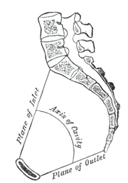
[19] The pelvic inclination angle is the single most important element of the human body posture and is adjusted at the hips.
The iliolumbar ligament passes between the tip of the transverse process of the fifth lumbar vertebra and the posterior part of the iliac crest.
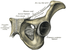
[20] The three extracapsular ligaments of the hip joint—the iliofemoral, ischiofemoral, and pubofemoral ligaments—form a twisting mechanism encircling the neck of the femur.
It can be thought of as the lower border of the thoracolumbar fascia and is occasionally accompanied by a smaller ligamentous band passing between the fourth lumbar vertebra and the iliac crest.
[26] The inferior parts of latissimus dorsi, one of the muscles of the upper limb, arises from the posterior third of the iliac crest.
[28] When the arm is adducted, latissimus dorsi can pull it backward and medially until the back of the hand covers the buttocks.
[27] In a longitudinal osteofibrous canal on either side of the spine there is a group of muscles called the erector spinae which is subdivided into a lateral superficial and a medial deep tract.
In the lateral tract, the iliocostalis lumborum and longissimus thoracis originates on the back of the sacrum and the posterior part of the iliac crest.
In the medial superficial group, on both sides of the centre of the abdominal wall (the linea alba), the rectus abdominis stretches from the cartilages of ribs V-VII and the sternum down to the pubic crest.
Quadratus lumborum arises from the posterior part of the iliac crest and extends to the rib XII and lumbar vertebrae I–IV.
The iliacus originates on the iliac fossa to join psoas at the iliopubic eminence to form the iliopsoas which is inserted into the lesser trochanter.
[32] The tensor fasciae latae arises on the anterior superior iliac spine and inserts into the iliotibial tract.
The thigh adductors have their origins on the inferior ramus of the pubic bone and are, with the exception of gracilis, inserted along the femoral shaft.
Three of the four muscles have their origins on the femur, while rectus femoris arises from the anterior inferior iliac spine and is thus the only of the four acting on two joints.
[36] The posterior thigh muscles have their origins on the inferior ischial ramus, with the exception of the short head of the biceps femoris.
[40][41] As the end of pregnancy approaches, the ligaments of the sacroiliac joint loosen, letting the pelvis outlet widen somewhat; this is easily noticeable in the cow.
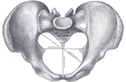
Throughout the 20th century pelvimetric measurements were made on pregnant women to determine whether a natural birth would be possible, a practice today limited to cases where a specific problem is suspected or following a caesarean delivery.
In 1933 and 1934 they published their typology, including the Greek names since then frequently quoted in various handbooks: Gynaecoid (gyne, woman), anthropoid (anthropos, human being), platypelloid (platys, flat), and android (aner, man).
[44][45] However, Caldwell and Moloy then complicated this simple fourfold scheme by dividing the pelvic inlet into posterior and anterior segments.
The dimensions of the head of the fetus and of the birth canal are accurately measured and compared, and the feasibility of labor can be predicted.
The pelvic girdle was present in early vertebrates, and can be tracked back to the paired fins of fish that were some of the earliest chordates.
The lengths of the ilium and ischium and their angles relative to the acetabulum are functionally important as they determine the moment arms for the hip extensor muscles that provide momentum during locomotion.
[53] In addition to this, the relatively wide shape (front to back) of the pelvis provides greater leverage for the gluteus medius and minimus.
In primates, the pelvis consists of four parts - the left and the right hip bones which meet in the mid-line ventrally and are fixed to the sacrum dorsally and the coccyx.
[55] The drying of the environment of East Africa in the period since the creation of the Red Sea and the African Rift Valley saw open woodlands replace the previous closed canopy forest.

2–4. Hip bone ( os coxae )
1. Sacrum ( os sacrum ), 2. Ilium ( os ilium ), 3. Ischium ( os ischii )
4. Pubic bone ( os pubis ) ( 4a. corpus , 4b. ramus superior , 4c. ramus inferior , 4d. tuberculum pubicum )
5. Pubic symphysis , 6. Acetabulum (of the hip joint ), 7. Obturator foramen , 8. Coccyx /tailbone ( os coccygis )
Dotted. Linea terminalis of the pelvic brim .