Hip bone
[3] The ischium forms the lower and back part of the hip bone and is located below the ilium and behind the pubis.
[4] The false pelvis is that portion superior to the pelvic brim; it is bounded by the alae of the ilia laterally and the sacral promontory and lumbar vertebrae posteriorly.
The hip bone is ossified from eight centers: three primary, one each for the ilium, ischium, and pubis, and five secondary, one each for the iliac crest, the anterior inferior spine (said to occur more frequently in the male than in the female), the tuberosity of the ischium, the pubic symphysis (more frequent in the female than in the male), and one or more for the Y-shaped piece at the bottom of the acetabulum.
The centers appear in the following order: in the lower part of the ilium, immediately above the greater sciatic notch, about the eighth or ninth week of fetal life; in the superior ramus of the ischium, about the third month; in the superior ramus of the pubis, between the fourth and fifth months.
At birth, the three primary centers are quite separate, the crest, the bottom of the acetabulum, the ischial tuberosity, and the inferior rami of the ischium and pubis being still cartilaginous.
By the seventh or eighth year, the inferior rami of the pubis and ischium are almost completely united by bone.
At about the age of puberty, ossification takes place in each of the remaining portions, and they join with the rest of the bone between the twentieth and twenty-fifth years.
Separate centers are frequently found for the pubic tubercle and the ischial spine, and for the crest and angle of the pubis.
Pelvimetry is the assessment of the female pelvis[7] in relation to the birth of a baby in order to detect an increased risk for obstructed labor.
The hip bones on each side usually connect with each other at the forward end, and are even solidly fused in lungfishes and sharks, but they never attach to the vertebral column.
[8] In practice, modern amphibians and reptiles have substantially modified this ancestral structure, based on their varied forms and lifestyles.
In birds, the pubic symphysis is present only in the ostrich, and the two hip bones are usually widely separated, making it easier to lay large eggs.
The same pattern is seen in all modern mammals, and the thyroid fenestra and obturator foramen have merged to form a single space.
The ilium is typically narrow and triangular in mammals, but is much larger in ungulates and humans, in which it anchors powerful gluteal muscles.

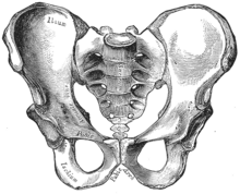
2–4. Hip bone ( os coxae )
1. Sacrum ( os sacrum ), 2. Ilium ( os ilium ), 3. Ischium ( os ischii )
4. Pubic bone ( os pubis ) ( 4a. corpus , 4b. ramus superior , 4c. ramus inferior , 4d. tuberculum pubicum )
5. Pubic symphysis , 6. Acetabulum (of the hip joint ), 7. Obturator foramen , 8. Coccyx /tailbone ( os coccygis )
Dotted. Linea terminalis of the pelvic brim .