Photoreceptor cell
The two classic photoreceptor cells are rods and cones, each contributing information used by the visual system to form an image of the environment, sight.
[2] These cells are thought not to contribute to sight directly, but have a role in the entrainment of the circadian rhythm and the pupillary reflex.
Closest to the visual field (and farthest from the brain) is the axon terminal, which releases a neurotransmitter called glutamate to bipolar cells.
Finally, closest to the brain (and farthest from the field of view) is the outer segment, the part of the photoreceptor that absorbs light.
Outer segments are actually modified cilia[5][6] that contain disks filled with opsin, the molecule that absorbs photons, as well as voltage-gated sodium channels.
In cone cells, there are different types of opsins that combine with retinal to form pigments called photopsins.
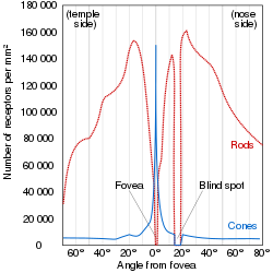
No photoreceptors are found at the blind spot, the area where ganglion cell fibers are collected into the optic nerve and leave the eye.
[9] The distribution of cone classes (L, M, S) are also nonhomogenous, with no S-cones in the fovea, and the ratio of L-cones to M-cones differing between individuals.
This polarization ultimately leads to either the transmittance or inhibition of a neural signal that will be fed to the brain via the optic nerve.
In the dark, cells have a relatively high concentration of cyclic guanosine 3'-5' monophosphate (cGMP), which opens cGMP-gated ion channels.
The movement of these positively charged ions into the cell (driven by their respective electrochemical gradient) depolarizes the membrane, and leads to the release of the neurotransmitter glutamate.
Unstimulated (in the dark), cyclic-nucleotide gated channels in the outer segment are open because cyclic GMP (cGMP) is bound to them.
[8] The rod and cone photoreceptors signal their absorption of photons via a decrease in the release of the neurotransmitter glutamate to bipolar cells at its axon terminal.
Absorption of a photon will hyperpolarize the photoreceptor and therefore result in the release of less glutamate at the presynaptic terminal to the bipolar cell.
This difference has important functional consequences: Comparison of human rod and cone cells, from Eric Kandel et al. in Principles of Neural Science.
There are five steps to developing photoreceptors: proliferation of multi-potent retinal progenitor cells (RPCs); restriction of competence of RPCs; cell fate specification; photoreceptor gene expression; and lastly axonal growth, synapse formation and outer segment growth.
Photoreceptor precursors come about through inhibition of Notch signaling and increased activity of various factors including achaete-scute homologue 1.
[13] In humans the ipRGCs contribute to non-image-forming functions like circadian rhythms, behavior and pupillary light reflex.
Birds have photoactive cerebrospinal fluid (CSF)-contacting neurons within the paraventricular organ that respond to light in the absence of input from the eyes or neurotransmitters.