Plane-form enamel hypoplasia
Plane-form enamel hypoplasia can be caused by a variety of factors, including severe illness/malnutrition, as well as specific conditions such as amelogenesis imperfecta and congenital syphilis.
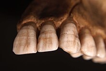
In severe cases enamel can be completely missing from areas of the crown, exposing the underlying dentine.
[3] Signs of plane-form enamel hypoplasia can be observed through pitting, depressions, and grooves seen on the surface of the teeth.
It is common for the disease to occur during the developmental stages of the teeth, and childhood illnesses, such as respiratory infections, are often linked to disturbance of the enamel formation.
Health conditions such as maternal malnutrition, infections, and exposure to harmful substances during pregnancy have been linked to enamel deficiency in the fetus.
[6] Preventative measures include limiting exposure to harmful chemicals, maintaining a nutrient-rich diet, and attending regular checkups during pregnancy.
Diagnosis typically beings with a visual examination, with the dentist looking for common signs such as pitting, depressions, and grooves on the tooth surface.
[11][10][5] Although prognosis information is limited, it has been found that Plane-form enamel hypoplasia is common within children, due to the tooth development being crucial during these stages of life.
Plane-form enamel hypoplasia is prevalent in regions that have limited access to dental care and poor nutritional resources.
As stated previously, nutritional deficiencies, systematic illnesses, infections, and exposure to toxins during tooth development can disturb the formation of the enamel.
Maternal smoking and certain medications can cause disruption during the pregnancy, leading to lack of enamel development in the fetus.
In 2022 research was conducted on 7,159 individuals within a multiethnic cohort, identifying genetic loci that is associated with Plane-form enamel hypoplasia.
The BMP2K and SLC4AR gene was located suggesting its contribution within tooth development, these findings highlight the complexity of enamel hypoplasia.
They found that using the microscope provided essential viewing that is needed to properly see enamel defects, suggesting that it would be valuable to use within routine dental practices.