Portal vein
Approximately 75% of total liver blood flow is through the portal vein, with the remainder coming from the hepatic artery proper.
In abdominal obesity fats, inflammatory cytokines and other toxic substances are transported by the portal vein from visceral fat into the liver, leading to hepatic insulin resistance and metabolic dysfunction–associated steatotic liver disease.
[1][2] Measuring approximately 8 cm (3 inches) long in adults,[3] the portal vein is located in the right upper quadrant of the abdomen, originating behind the neck of the pancreas.
These vessels ultimately empty into the hepatic sinusoids to supply blood to the liver.
In cases of portal hypertension these anastomoses may become engorged, dilated, or varicosed and subsequently rupture.
[4] The portal vein and hepatic arteries form the liver's dual blood supply.
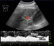
[8] On Doppler ultrasonography, the main portal vein (MPV) peak systolic velocity normally ranges between 20 cm/s and 40 cm/s.
[9] Clinical signs of portal hypertension include those of chronic liver disease: ascites, esophageal varices, spider nevi, caput medusae, and palmar erythema.
[9] Portal vein pulsatility can be quantified by pulsatility indices (PI), where an index above a certain cutoff indicates pathology: Pylephlebitis is infection of the portal vein, usually arising from an infectious intra-abdominal process such as diverticulitis.