Horse hoof
The upper, almost circular limit of the hoof capsule is the coronet (also called coronary band), which is at an angle to the ground of roughly similar magnitude in each pair of feet (i.e., fronts and backs).
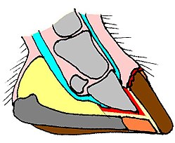
At the heels, the palmar/plantar portions of the walls bend inward sharply, following the external surface of the collateral grooves to form the bars.
The lower surface of the hoof, from the outer walls and the inner frog and bars, is covered by an exfoliating keratinised material, called the 'sole'.
The walls are considered as a protective shield covering the sensitive internal hoof tissues (like the exoskeleton of arthropods), as a structure devoted to dissipating the energy of concussion, and as a surface to provide grip on different terrains.
The water line's thickness increases proportionally to the distance from the coronet and, in the lower third of the walls, is thicker than the pigmented layer.
It is softer and fibrous in structure and light in color; white in a freshly trimmed hoof, yellowish or gray after exposure to air and dirt.
If the wall does not wear naturally from sufficient movement on abrasive terrain, then it will protrude from the solar surface.
It is dark gray-blackish in color and of a rubbery consistency, suggesting its role as a shock absorber and grip tool on hard, smooth ground.
The frog also acts like a pump to move the blood back to the heart, a great distance from the relatively thin leg to the main organ of the circulatory system.
In the stabled horse, the frog does not wear but degrades, due to bacterial and fungal activity, to an irregular, soft, slashed surface.
[citation needed] For good health, the horse requires dry areas to stand.
If exposed to constant wet or damp environments, the frog can develop a bacterial infection called thrush.
If there is no contact, as in shod hooves or when the walls are too long or the movement poor, the lower surface of the sole has a crumbly consistency, and it is easily abraded by scratching it with a hoofpick.
It is often caused by a horse treading on a stone or sharp type of object, landings from high jumps and excessive exposure to snow.
The corium, a dermo-epidermal, highly vascularized and innervated layer between the wall and the coffin bone, has a parallel, laminar shape, and is named the laminae.
In the adult horse, it develops a fibrocartilaginous network that helps support the bony column.
Normal transformation of the digital cushion into fibrocartilagineous tissue is now considered a key goal, both for prevention of, and for rehabilitation of recovering cases of navicular syndrome.
Hooves are a plastic structure and their time-related, very complex changes can be considered in the short term (days/weeks) and over the horse's lifespan.
The resulting 'dead' superficial layer serves a protective function, saving underlying living tissues from injury, from dehydration, and from fungal and bacterial attack.
The wall does not exfoliate at all; it is constantly growing downward (about 1 cm per month), and under normal circumstances self-trims by wearing or chipping by ground contact.
In wild and feral horses, solar, frog and periople materials grow outwards and exfoliate at the surface by ground contact and wearing.
In the domesticated horse, movement and typical ground hardness are insufficient to allow self-trimming, so humans have to care for them by trimming the walls and the frog, and scraping off the dead sole.
The front and hind hooves are identical in the foal but differ visibly in the adult horse.
The resulting conformation allows a heavy, strong body to move with high speed on any ground, and most efficiently on open, hard, flat areas like prairies and deserts (an example of cursorial specialisation).
Quittor, an infection of collateral cartilages in the lower leg is also sometimes seen, although most commonly in draft horses.