Role of skin in locomotion
Soft bodied animals such as starfish rely on the arrangement of the fibers in their tube feet for movement.
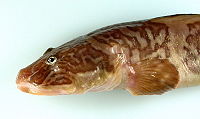
Eels, snakes, and fish use their skin like an external tendon to generate the propulsive forces need for undulatory locomotion.
The term "Soft Bodied" refers to animals which lack typical systems of skeletal support - included in these are most insect larvae and true worms.
Internal to the patterned fiber layer is typically a liquid filled cavity, which is used to generate hydrostatic pressures for movement.
[1] Some animals that exhibit soft bodied locomotion include starfish, octopus, and flatworms.
[2] This structure plays a role in invertebrate support and locomotor systems and is used for the tube feet in starfish and body of worms.
[4] The arrangement of connective tissue fibers determines the range of motion of a body, and serves as an antagonist against muscle contraction.
This cross helical arrangement is seen in the tube feet starfish, different types of worms and suckers in octopus.
This cross helical arrangement allows for the connective tissue layers to evenly distribute force throughout the hydrostatic body.
This arrangement of connective tissue fibers creates a stiffer body wall and more muscle antagonism, which allows for more elastic force to be generated and released during movement.
[7] In an eel with the cross helical fiber arrangement, muscle contraction in the anterior region bends the fish, and so the skin on the convex side is extended in the longitudinal direction.
[7] The skin act like an external tendon allowing for an eel to generate a greater propulsive force per muscle contraction.
When the dermis is placed in tension, and resistance to bending is developed, which is referred to as flexural stiffness of the fish skin.
One of the interesting aspects of snakeskin are folds of intersquamous skin between longitudinally oriented scale rows.
[9] The function of these folds is to permit the circumference of the snake to increase, allowing prey to pass into the stomach during feeling.
[10] Differences in the local dermal structures, such as variations in the diameters and orientation of collagen fibers within the intersquamous skin create local differences in the mechanical properties of the snake skin, thus allowing it to adapt to the stresses and strains during the feeding process.
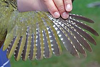
Avain skin must be structurally arranged such that "the coat of feathers" remains smooth and intact during flight.
Within the dermal and epidermal layer of bat wing skin, the connective tissue and muscle fibers provide the structural support.
Of the muscles that insert themselves into the mesh scaffolding, larger muscles anchor the skin to the bone and control the membrane tension and camber of the bat wing during flight,[14] whereas smaller muscles, which originate from within the mesh scaffolding, attach to collagen fibers within the fiber network and modulate bone loading and allow for precise control of wing shape and tension.
Within the mesh scaffolding of bat wing skin, collagen fibers cross bones perpendicular to the long axes of the bones, therefore mechanical properties of bat wing skin oriented perpendicular to the long axes of the bones exhibit a lower stiffness than the skin that is oriented parallel to the long axes of the bodies.
Flexible skin is necessary for the direction perpendicular to the long axes of the bones for facilitating the shape changes needed for movement and control during flight.
[14] This anisotropy of bat wing skin is also useful as method of storing and releasing elastic energy, particularly during the downstroke.
The skeletal supports and muscle erect the flight membrane and control the gliding using the patagia.
A large portion of the dorsal scales of the patagia are arranged in regular rib-like pattern, which guide the flow of air and allow for the lizard to behave as an airfoil.
To better understand the structure of avian skin, avian skin has been broken down into three different functional components: This functional component consists of the only of the structural features of the feather tracts, namely the cutis, and connective tissue layer fascia superficialis.
The smooth muscles of the apteria counteract the horizontal forces experienced by the feather follicles.