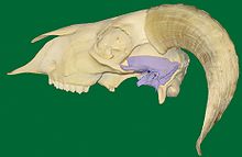
Temporomandibular joint
The anterior portion of the disc serves as the insertion site for the superior head of the lateral pterygoid.
[2] The central area of the disc is avascular and lacks innervation, thus getting its nutrients from the surrounding synovial fluid.
With age, the entire disc thins and may undergo the addition of cartilage in the central part, changes that may lead to impaired movement of the joint.
[3] The lower joint compartment formed by the mandible and the articular disc is involved in rotational movement—this is the initial movement of the jaw when the mouth opens.
The upper joint compartment formed by the articular disc and the temporal bone is involved in translational movement—this is the secondary gliding motion of the jaw as it is opened widely.
[2] In some cases of anterior disc displacement, the pain felt during movement of the mandible is due to the condyle compressing this area against the articular surface of the temporal bone.
[citation needed] Free nerve endings, many of which act as nociceptors, innervate the bones, ligaments, and muscles of the TMJ.
[11] At approximately 10 weeks the component of the fetus future joint becomes evident in the mesenchyme between condylar cartilage of the mandible and the developing temporal bone.
The developing superior head of the lateral pterygoid muscle attaches to the anterior portion of the fetal disk.
This growth center consists of hyaline cartilage underneath the periosteum on the articulating surface of the condyle.
Instead, the muscular balance and proprioceptive feedback allow a physiologic rest for the mandible, an interocclusal clearance, or freeway space, which is 2 to 4 mm between the teeth.
This is accomplished by translation of the condyle down the articular eminence (in the upper portion of the joint) without any more than the slightest amount of rotation taking place (in the lower portion of the joint), other than that necessary to allow the mandibular incisors to come in front of the maxillary incisors without running into them.
The latter terms, although a bit outdated, are actually more precise, as they define the sides by the movements of the respective condyles.
These four muscles, all innervated by V3, or the mandibular division of the trigeminal nerve, work in different groups to move the mandible in different directions.
This term is used to refer to a group of problems involving the temporomandibular joints and the muscles, tendons, ligaments, blood vessels, and other tissues associated with them.
Although rare, other pathologic conditions may also affect the function of temporomandibular joints, causing pain and swelling.
These conditions include chondrosarcoma, osteosarcoma, giant cell tumor, and aneurysmal bone cyst.
This, as explained above, is usually very painful, because, unlike these adjacent tissues, the central portion of the disc contains no sensory innervation.
On opening, a "pop" or "click" can sometimes be heard and usually felt also, indicating the condyle is moving back onto the disk, known as "reducing the joint" (disc displacement with reduction).
Upon clenching, the condyle compresses the bilaminar area, and the nerves, arteries, and veins against the temporal fossa, causing pain and inflammation.