Tetramer assay
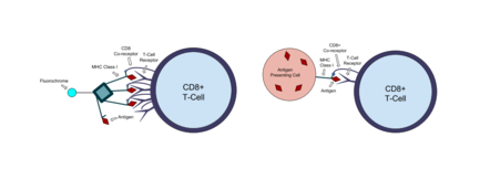
[1] The tetramers used in the assay are made up of four major histocompatibility complex (MHC) molecules, which are found on the surface of most cells in the body.
[2] MHC molecules present peptides to T-cells as a way to communicate the presence of viruses, bacteria, cancerous mutations, or other antigens in a cell.
[2][3] Tetramer stains allow for the visualization, quantification, and sorting of these cells by flow cytometry, which is extremely useful in immunology.
Tetramer stains can also be paired with functional assays like ELIspot, which detects the number of cytokine secreting cells in a sample.
Class I MHC molecules are made up of a polymorphic heavy α-chain associated with an invariant light chain beta-2 microglobulin (β2m).
Highly active formulations of a broad range of MHC class I molecules[10] now allows non-experts users to make their own custom peptide-MHC complexes from day-to-day in any lab without special equipment.
Class II MHC molecules present extracellular antigens, allowing helper T-cells to detect bacteria,[11] fungi, and parasites.
This was compared to results of cytotoxic assays and plasma RNA viral load to characterize the function of CTLs in HIV infection.
This allowed for comparison of the immune response (the number of T-cells that target a virus) in two different vaccine delivery methods.