Femur
On average, the femur length accounts for 26.74% of a person's height,[4] a ratio found in both men and women across most ethnic groups with minimal variation.
[3] The transition area between the head and neck is quite rough due to attachment of muscles and the hip joint capsule.
The highest point of the greater trochanter is located higher than the collum and reaches the midpoint of the hip joint.
The trochanteric fossa is a deep depression bounded posteriorly by the intertrochanteric crest on the medial surface of the greater trochanter.
[3] The third trochanter is a bony projection occasionally present on the proximal femur near the superior border of the gluteal tuberosity.
[6] A structure of minor importance in humans, the incidence of the third trochanter varies from 17–72% between ethnic groups and it is frequently reported as more common in females than in males.
[3] Anteriorly, the condyles are slightly prominent and are separated by a smooth shallow articular depression called the patellar surface.
The medial condyle is the longer and, when the femur is held with its body perpendicular, projects to a lower level.
When, however, the femur is in its natural oblique position the lower surfaces of the two condyles lie practically in the same horizontal plane.
The condyles are not quite parallel with one another; the long axis of the lateral is almost directly antero-posterior, but that of the medial runs backward and medialward.
This fossa is limited above by a ridge, the intercondyloid line, and below by the central part of the posterior margin of the patellar surface.
As the femur is the only bone in the thigh, it serves as an attachment point for all the muscles that exert their force over the hip and knee joints.
The neck of the femur is generally minimal or absent in the most primitive forms, reflecting a simple attachment to the acetabulum.
The greater trochanter was present in the extinct archosaurs, as well as in modern birds and mammals, being associated with the loss of the primitive sprawling gait.
In some snakes, the protruding end of a pelvic spur, a vestigial pelvis and femur remnant which is not connected to the rest of the skeleton, plays a role in mating.
This role in mating is hypothesized to have possibly occurred in Basilosauridae, an extinct family of whales with well-defined femurs, lower legs and feet.
Occasionally, the genes that code for longer extremities cause a modern whale to develop miniature legs (atavism).
[12] One of the earliest known vertebrates to have a femur is the Eusthenopteron, a prehistoric lobe-finned fish from the Late Devonian period.
A recent study has revealed that bone is a significantly richer source of persistent DNA viruses than previously thought.
The usage is not homologous with that of vertebrate anatomy; the term "femur" simply has been adopted by analogy and refers, where applicable, to the most proximal of (usually) the two longest jointed segments of the legs of the Arthropoda.
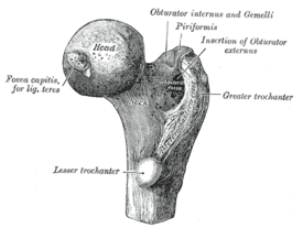


(seen from the front)

(seen from the back)
