Human tooth development
Tooth development or odontogenesis is the complex process by which teeth form from embryonic cells, grow, and erupt into the mouth.
[2] Additionally, the junction between the dental papilla and inner enamel epithelium determines the crown shape of a tooth.
The dental sac or follicle gives rise to three important entities: cementoblasts, osteoblasts, and fibroblasts.
All stages (bud, cap, bell, crown), growth and morphogenesis of the teeth are regulated by a protein called sonic hedgehog.
The dental lamina connects the developing tooth bud to the epithelial layer of the mouth for a significant time.
The dental organ is bell-shaped during this stage, and the majority of its cells are called stellate reticulum because of their star-shaped appearance.
The dental lamina disintegrates, leaving the developing teeth completely separated from the epithelium of the oral cavity; the two will not join again until the final eruption of the tooth into the mouth.
The components for particular types of teeth, such as incisors, are localized in one area and dissipate rapidly in different parts of the mouth.
[citation needed] The other dominant hypothesis, the "clone model", proposes that the epithelium programs a group of ectomesenchymal cells to generate teeth of particular shapes.
Nasmyth membrane then easily picks up stain from food debris and is hard to remove except by selective polishing.
Odontoblasts increase in size, eliminating the availability of any extracellular resources to contribute to an organic matrix for mineralization.
Additionally, the larger odontoblasts cause collagen to be secreted in smaller amounts, which results in more tightly arranged, heterogeneous nucleation that is used for mineralization.
As mineralization takes place, the cementoblasts move away from the cementum, and the fibers left along the surface eventually join the forming periodontal ligaments.
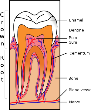
[citation needed] The periodontium, which is the supporting structure of a tooth, consists of the cementum, periodontal ligaments, gingiva, and alveolar bone.
Alveolar bone surrounds the roots of teeth to provide support and creates what is commonly called a "socket".
Periodontal ligaments connect the alveolar bone to the cementum, and the gingiva is the surrounding tissue visible in the mouth.
Specific events leading to the formation of the periodontal ligament vary between deciduous (baby) and permanent teeth and among various species of animals.
[36] Similar to the formation of primary cementum, collagen fibers are created on the surface nearest the tooth, and they remain there until attaching to periodontal ligaments.
This results in the perpetually increasing size of the junctional epithelial layer and the isolation of the remnants of ameloblasts from any source of nutrition.
[citation needed] Frequently, nerves and blood vessels run parallel to each other in the body, and the formation of both usually takes place simultaneously and in a similar fashion.
The number of blood vessels reaches a maximum at the beginning of the crown stage, and the dental papilla eventually forms in the pulp of a tooth.
This theory postulated that a ligament below a tooth, which Sicher observed under a microscope on a histologic slide, was responsible for eruption.
[41] The most widely held current theory is that while several forces might be involved in eruption, the periodontal ligaments provide the main impetus for the process.
Theorists hypothesize that the periodontal ligaments promote eruption through the shrinking and cross-linking of their collagen fibers and the contraction of their fibroblasts.
The dental theory is the low levels of fluoride incorporation and very mild fluorosis makes the tooth more resistant to demineralization and subsequent decay.
[61][62][63] Bisphenol A (BPA) is a hormone-disrupting chemical that has been implicated in having negative effects on human health, including, but not limited to, fetal development.
As shown in animal studies which mimic human enamel, the mother's consumption of products with BPA during pregnancy can lead to the child's tooth development being obstructed.
[65] About 86% of these cases involve a single extra tooth in the mouth, most commonly found in the maxilla, where the incisors are located.
[69] Some systemic conditions may cause delayed tooth development, such as nutritional factors, endocrine disorders (hypothyroidism, hypopituitarism, hypoparathyroidism, pseudohypoparathyroidism),[70] undiagnosed and untreated celiac disease,[70][71] anemia, prematurity, low birth weight, renal failure, heavy metal intoxication or tobacco smoke, among others.
[72] Teeth affected by regional odontodysplasia nevAmelogenesis imperfecta is an autosomal dominant disease characterized by a defect in dental enamel formation.


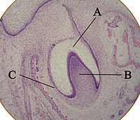
A: enamel organ
B: dental papilla
C: dental follicle


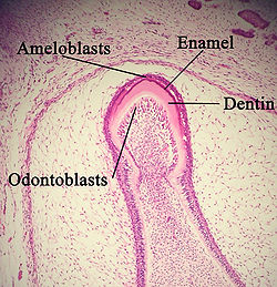

A: enamel
B: dentin

A: dentin
B: cementum

A: tooth
B: gingiva
C: bone
D: periodontal ligaments