Toothcomb
The toothcomb occurs in lemuriform primates (which include lemurs and lorisoids), treeshrews, colugos, hyraxes, and some African antelopes.
The toothcombs of colugos and hyraxes take a different form with the individual incisors being serrated, providing multiple tines per tooth.
The lack of a distinct toothcomb in the fossil record before to 40 mya has created a conflict with molecular clock studies that suggest an older divergence between lemurs and lorisoids, and the existence of a ghost lineage of lemuriform primates in Africa.
[6] Toothcombs can also be found in colugos and treeshrews, both close relatives of primates; however, the structures are different and these are considered examples of convergent evolution.
[7][8] Likewise, small- or medium-sized African antelopes, such as the impala (Aepyceros melampus),[9] have a similar structure sometimes referred to as the "lateral dental grooming apparatus".
[10][11] Living and extinct hyraxes (hyracoids) also exhibit a toothcomb, although the number of tines in the comb varies throughout the fossil record.
[12] Dating to the Eocene epoch over 50 mya, Chriacus and Thryptacodon—two types of arctocyonids (primitive placental mammals)—also possessed an independently evolved toothcomb.
[15] The French anatomist Henri Marie Ducrotay de Blainville first identified the two lateral teeth of the lemuriform toothcomb as canines in 1840.
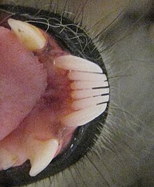
[15] The lemuriform toothcomb is kept clean by the sublingua or "under-tongue", a specialized muscular structure that acts like a toothbrush to remove hair and other debris.
The sublingua can extend below the end of the tongue and is tipped with keratinized, serrated points that rake between the front teeth.
[28] The aye-aye also lost its toothcomb, replacing it with continually growing (hypselodont) front teeth, similar to the incisors of rodents.
[10] As a homologous structure in lemuriforms, the toothcomb serves variable biological roles, despite its superficially stereotypic shape and appearance.
[31] More than 100 years later, the grooming function was questioned since it was difficult to observe and the interdental spaces were thought to be too small for fur.
[14] Among non-primates, the extinct Chriacus exhibits microscopic groves on its toothcomb,[14] but the Philippine colugo (Cynocephalus volans) does not.
[10][11] In lemuriform primates, the toothcomb may also play a secondary role in olfaction, which may account for the size reduction of the poorly studied upper incisors.
[42] Furthermore, the size reduction of the upper incisors may create a gap between the teeth (interincisal diastema) that connects the philtrum (a cleft in the middle of the wet nose, or rhinarium) to the vomeronasal organ in the roof of the mouth.
[14][44] In fork-marked lemurs, the toothcomb is specially adapted to minimize food trapment since the interdental spaces are greatly reduced.
[46] The more robust structure of their toothcomb is thought to help it withstand the compressive forces experienced during regular bark-prising.
[34] However, the inclusion of the canines into the toothcomb must have required exceptional conditions, since large lemuriforms have secondarily modified caniniform premolars to substitute for the loss.
[52] In some adapids, the crests of the lower incisors and canines align to form functional cropping unit, and the American paleontologist Philip D. Gingerich has suggested this foreshadowed the development of the lemuriform toothcomb.
[56] Stem lemuriforms, including Djebelemur and 'Anchomomys' milleri, have been found in Africa and date from 50 to 48 mya and were very distinct from European adapiforms.
[c][60] Based on large, procumbent lower teeth, Plesiopithecus, a fossil primate found in late Eocene deposits at the Fayum Depression in Egypt, is thought to be most closely related to lemuriforms.
[49] This would conflict with the molecular clock estimates by evolutionary anthropologist Anne Yoder and others, which predict lemur–lorisoid divergence dating between 61 and 90.8 mya.
[72] Also the interdental spaces seen in most lemuriforms favor fur combing and would also promote bacterial growth and tooth decay if used for exudate feeding.