White matter dissection
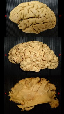
White matter dissection refers to a special anatomical technique able to reveal the subcortical organization of white matter fibers in the human or animal cadaver brain.
The first studies of cerebral white matter (WM) were described by Galen and by the subsequent efforts of Vesalius on human cadaver specimens.
[2][3] The interest for the deep anatomy of the brain pushed anatomist during centuries to create and develop different techniques for specimen preparation and dissection in order to better reveal the complex white matter architectural organization.
This process made possible to peel off the cortex from the brain surface without damaging the subcortical white matter organization underneath.
[4][8][9] White matter fibre dissection is nowadays considered as a valuable tool to enhance our knowledge about brain connectivity,[5][9][10][1] and has been used to validate tractographic results and vice versa with good consistency between the two techniques,[11] but also for neurosurgical training and neuroanatomical teaching.