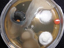
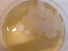
Agar plate
At some point during a successful "streak", the number of organisms deposited will be such that distinct individual colonies will grow in that area which may be removed for further culturing, using another sterile loop.
This type of analysis is often used to check the viability of cells and is performed with pinners (often also called froggers).
In this technique, cells are grown in a liquid culture, in which a small volume is pipetted on the agar plate and then spread out with the beads.
[3] Like other growth media, the formulations of agar used in plates may be classified as either "defined" or "undefined"; a defined medium is synthesized from individual chemicals required by the organism so the exact molecular composition is known, whereas an undefined medium is made from natural products such as yeast extract, where the precise composition is unknown.
This correlates to some degree with defined and undefined media; undefined media, made from natural products and containing an unknown combination of very many organic molecules, is typically more permissive in terms of supplying the needs of a wider variety of organisms.
BAPs are enriched, and differential media is used to isolate fastidious organisms and detect hemolytic activity.
β-Hemolytic activity will show lysis and complete digestion of red blood cell contents surrounding a colony.