Anatomy
In recent years, there has been a significant increase in the use of advanced imaging techniques, such as MRI and CT scans, which allow for more detailed and accurate visualizations of the body's structures.
They have an internal digestive chamber, with one or two openings; the gametes are produced in multicellular sex organs, and the zygotes include a blastula stage in their embryonic development.
In more complex animals, specialized receptor cells such as chemoreceptors and photoreceptors are found in groups and send messages along neural networks to other parts of the organism.
Low frequency vibrations are detected by the lateral line system of sense organs that run along the length of the sides of fish, and these respond to nearby movements and to changes in water pressure.
Salamanders resemble lizards in appearance; their short legs project sideways, the belly is close to or in contact with the ground and they have a long tail.
Reptiles are unable to use their skin for respiration as do amphibians and have a more efficient respiratory system drawing air into their lungs by expanding their chest walls.
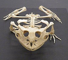
The skeleton consists of a skull, a hyoid bone, spine and ribs though a few species retain a vestige of the pelvis and rear limbs in the form of pelvic spurs.
Their forked tongues are used as organs of taste and smell and some species have sensory pits on their heads enabling them to locate warm-blooded prey.
[47] Invertebrates constitute a vast array of living organisms ranging from the simplest unicellular eukaryotes such as Paramecium to such complex multicellular animals as the octopus, lobster and dragonfly.
Locomotion is often provided by cilia or flagella or may proceed via the advance of pseudopodia, food may be gathered by phagocytosis, energy needs may be supplied by photosynthesis and the cell may be supported by an endoskeleton or an exoskeleton.
Calcium carbonate constitutes the shells of molluscs, brachiopods and some tube-building polychaete worms and silica forms the exoskeleton of the microscopic diatoms and radiolaria.
In 1600 BCE, the Edwin Smith Papyrus, an Ancient Egyptian medical text, described the heart and its vessels, as well as the brain and its meninges and cerebrospinal fluid, and the liver, spleen, kidneys, uterus and bladder.
Phenomenal anatomical observations of the human body were made, which contributed to the understanding of the brain, eye, liver, reproductive organs, and nervous system.
Great patronage of the arts and sciences from the Ptolemaic dynasty of Egypt helped raise Alexandria up, further rivalling other Greek states' cultural and scientific achievements.
[63] Some of the works included classifying the system of the pulse, the discovery that human arteries had thicker walls than veins, and that the atria were parts of the heart.
Herophilus's knowledge of the human body has provided vital input towards understanding the brain, eye, liver, reproductive organs, and nervous system and characterizing the course of the disease.
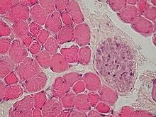
[68] The anatomy of the muscles and skeleton is described in the Hippocratic Corpus, an Ancient Greek medical work written by unknown authors.
Also in the 4th century BCE, Herophilos and Erasistratus produced more accurate anatomical descriptions based on vivisection of criminals in Alexandria during the Ptolemaic period.
[70][71] In the 2nd century, Galen of Pergamum, an anatomist, clinician, writer, and philosopher,[72] wrote the final and highly influential anatomy treatise of ancient times.
[75]: 120–121 Between 1275 and 1326, the anatomists Mondino de Luzzi, Alessandro Achillini and Antonio Benivieni at Bologna carried out the first systematic human dissections since ancient times.
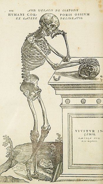
[81] The accurate and intricately detailed illustrations, often in allegorical poses against Italianate landscapes, are thought to have been made by the artist Jan van Calcar, a pupil of Titian.
Philadelphia, Baltimore, and New York were all renowned for body snatching activity as criminals raided graveyards at night, removing newly buried corpses from their coffins.
The practice was halted in Britain by the Anatomy Act of 1832,[86][87] while in the United States, similar legislation was enacted after the physician William S. Forbes of Jefferson Medical College was found guilty in 1882 of "complicity with resurrectionists in the despoliation of graves in Lebanon Cemetery".
As well as teaching, he collected many vertebrate skeletons for his museum of comparative anatomy, published over 70 research papers, and became famous for his public dissection of the Tay Whale.
Semmelweis showed that when the trainees washed their hands in chlorinated lime before each clinical examination, the incidence of puerperal fever among the mothers could be reduced dramatically.
[93] Before the modern medical era, the primary means for studying the internal structures of the body were dissection of the dead and inspection, palpation, and auscultation of the living.
Technical advances in the development of achromatic lenses increased the resolving power of the microscope, and around 1839, Matthias Jakob Schleiden and Theodor Schwann identified that cells were the fundamental unit of organization of all living things.
The study of small structures involved passing light through them, and the microtome was invented to provide sufficiently thin slices of tissue to examine.
The invention of the electron microscope brought a significant advance in resolution power and allowed research into the ultrastructure of cells and the organelles and other structures within them.
About the same time, in the 1950s, the use of X-ray diffraction for studying the crystal structures of proteins, nucleic acids, and other biological molecules gave rise to a new field of molecular anatomy.