Ascaris lumbricoides
An acute inflammatory reaction can occur if some of the worms get lost during this migration process and accumulate in other organs of the body.
One hypothesis to account for this behavior is that the migration mimics an intermediate host, which would be required for juveniles of an ancestral form to develop to the third stage.
Another possibility is that tissue migration enables faster growth and larger size, which increases reproductive capacity.
[2] While infection occurs throughout most of the world, ascariasis is most common in sub-Saharan Africa, the Americas, China, and east Asia.
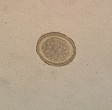
[9] Eggs of A. lumbricoides have been identified in coprolites in the Americas, Europe, Africa, the Middle East, and New Zealand, the oldest ones being more than 24,000 years old.
[14] Often, no symptoms are presented with a minor A. lumbricoides infection, the inevitable consequence being the e.g. once a year passage of such clearly visible worm(s) on close inspection.
[18] Ascaris lumbricoides is primarily distributed in tropical and subtropical regions around the world, particularly in areas with poor sanitation and hygiene practices.
It is most prevalent in sub-Saharan Africa, Southeast Asia (including countries like India, Bangladesh, and Indonesia), and parts of Latin America, where inadequate sanitation infrastructure and the use of human faeces as fertilizer contribute to its spread.
[19] Preventing any fecal-borne disease requires educated hygienic habits/culture and effective fecal treatment systems.
The eggs may get onto vegetables when improperly processed human feces of infected people are used as fertilizer for food crops.
In 1855, Ascaris eggs were found in human faeces by Henry Ransom in England then this was described in the literature two years later by Casimir-Joseph Davaine in France.
[24] Development was thought to occur directly within the bowel lumen but Francis Stewart in Hong Kong in 1916 fed eggs to rats, then later mice, and found infective larvae in the faeces and in the lungs but no mature worms.
In 1918, Sadao Yoshida ingested larvae recovered from the trachea of a guinea pig, then found eggs in his own stools 76 days later.