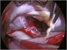
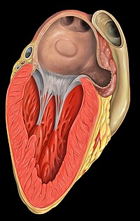
Mitral valve
The chordae tendineae are inelastic tendons attached at one end to papillary muscles in the left ventricle, and at the other to the valve cusps.
When the left ventricle contracts, the pressure in the ventricle forces the valve to close, while the tendons keep the leaflets coapting together and prevent the valve from opening in the wrong direction (thus preventing blood flowing back to the left atrium).
[11] The annulus contracts and reduces its surface area during systole to help provide complete closure of the leaflets.
Expansion of the annulus can result in leaflets that do not join soundly together, leading to functional mitral regurgitation.
Microscopically, there is no evidence of an annular structure anteriorly, where the mitral valve leaflet is contiguous with the posterior aortic root.
About 70 to 80% of the blood that travels across the mitral valve occurs during the early filling phase of the left ventricle.
This early filling phase is due to active relaxation of the ventricular myocardium, causing a pressure gradient that allows a rapid flow of blood from the left atrium, across the mitral valve.
This reduction in annulus size at the end of atrial systole may be important for the proper coapting of the leaflets of the mitral valve when the left ventricle contracts and pumps blood.
Classic mitral valve prolapse is caused by an excess of connective tissue that thickens the spongiosa layer of the cusp and separates collagen bundles in the fibrosa.
This weakens the cusps and adjacent tissue, resulting in an increased cuspal area and lengthening of the chordae tendineae.
Advanced lesions—also commonly involving the posterior leaflet—lead to leaflet folding, inversion, and displacement toward the left atrium.
[15] A valve prolapse can result in mitral insufficiency, which is the regurgitation or backflow of blood from the left ventricle to the left atrium due to the incomplete closure of the valve causing a systolic murmur heard at the apex of the heart.
This increase in pressure in the left atrium and pulmonary circuit can lead to symptoms like fatigue, shortness of breath, and atrial fibrillation over time.
[18] Rarely there can be a severe form of calcification of the mitral valve annulus that can be mistaken for an intracardiac mass or thrombus.
Common causes include, but is not limited to, Barlow disease, myxomatous degeneration, inflammation, and papillary muscle rupture.