Bivalve shell
The bivalve shell not only serves as protection from predators and physical damage, but also for adductor muscle attachment, which can allow the mollusc to "swim" short distances by flapping the valves.
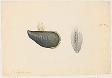
The shell is secreted by a soft part of the molluscan body known as the mantle and has several layers, typically made of calcium carbonate precipitated out into an organic matrix.
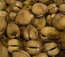
The shells of dead bivalves commonly wash up on beaches (often as separate valves) and along the edges of lakes, rivers and streams.
They are collected by professional and amateur conchologists and are sometimes harvested for commercial sale in the international shell trade or for use in glue, chalk, or varnish, occasionally to the detriment of the local ecology.
The mantle, a thin membrane surrounding the body, secretes the shell valves, ligament, and hinge teeth.
In some bivalves the mantle edges fuse to form siphons, which take in and expel water during suspension feeding.
The mechanical properties of bivalve shells and their relatedness to microstructure was first published in 1969 by Stephen Wainwright at Duke University.
For example, one type of bivalve, Cerastoderma edule, was studied with scanning electron microscopy (SEM) and nanoindentation to determine if exposure to higher levels of carbon dioxide would affect the structure of the shell.
Fortunately for the bivalves, there appeared to be no strong correlation between exposure to high carbon dioxide partial pressures and shell hardness.
[7] A general reader may believe that defects and non-uniformity would decrease the strength of the bivalve shell, but that is not necessarily the case.
For example, when a type of bivalve, Tridacna gigas, was modelled and analyzed, it was found to be highly oriented in along a singular axis.
Compression tests have revealed that the presence of those ridges allows for more resistance to fracture than those with polished edges.
The oldest point of a bivalve shell is called the beak, and the raised area around it is known as the umbo (plural umbones).
Similar annual pallial line scars on the interior of the valves are more easily seen in dark colored shells, but these may be overgrown and obscured by further deposition of hard material.
The most accurate but most time-consuming method is the microscopic examination of sections through the outer prismatic layer of the shell.
They are generally conservative within major groups, and have historically provided a convenient means upon which to base classification schemes and the phylogenetic order.


- Plane of symmetry
- Growth lines
- Ligament
- Umbo