Histology
[5] In medicine, histopathology is the branch of histology that includes the microscopic identification and study of diseased tissue.
[10] Trained physicians, frequently licensed pathologists, perform histopathological examination and provide diagnostic information based on their observations.
This process, while preserving the structural integrity of the cells and tissue can damage the biological functionality of proteins, particularly enzymes.
Formalin fixation leads to degradation of mRNA, miRNA, and DNA as well as denaturation and modification of proteins in tissues.
[9][5] In general, water must first be removed from tissues (dehydration) and replaced with a medium that either solidifies directly, or with an intermediary fluid (clearing) that is miscible with the embedding media.
[12][13] Paraffin is immiscible with water, the main constituent of biological tissue, so it must first be removed in a series of dehydration steps.
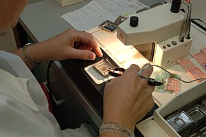
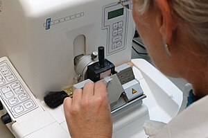
[9] In most histology, or histopathology laboratories the dehydration, clearing, and wax infiltration are carried out in tissue processors which automate this process.
[13][12] Paraffin wax does not always provide a sufficiently hard matrix for cutting very thin sections (which are especially important for electron microscopy).
Pre-frozen tissues are placed into molds with the liquid embedding material, usually a water-based glycol, OCT, TBS, Cryogen, or resin, which is then frozen to form hardened blocks.
[9] For transmission electron microscopy (TEM), a diamond or glass knife mounted in an ultramicrotome is used to cut between 50 and 150 nanometer thick tissue sections.
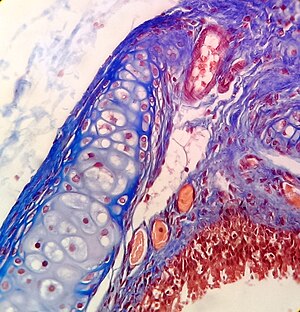
[12] A commonly performed histochemical technique that targets a specific chemical is the Perls' Prussian blue reaction, used to demonstrate iron deposits[12] in diseases like hemochromatosis.
More commonly, autoradiography is used in visualizing the locations to which a radioactive substance has been transported within the body, such as cells in S phase (undergoing DNA replication) which incorporate tritiated thymidine, or sites to which radiolabeled nucleic acid probes bind in in situ hybridization.
For autoradiography on a microscopic level, the slide is typically dipped into liquid nuclear tract emulsion, which dries to form the exposure film.
[9] Uranyl acetate and lead citrate are commonly used to impart contrast to tissue in the electron microscope.
Ultramicrotomy is a method of preparing extremely thin sections for transmission electron microscope (TEM) analysis.
While studying the structure of the lung, Malpighi noticed its membranous alveoli and the hair-like connections between veins and arteries, which he named capillaries.
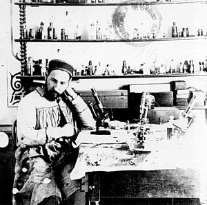
[28] The 1906 Nobel Prize in Physiology or Medicine was awarded to histologists Camillo Golgi and Santiago Ramon y Cajal.
Ramón y Cajal won the prize for his correct theory, and Golgi for the silver-staining technique that he invented to make it possible.