[5] During ventricular systole the ventricles contract and vigorously pulse (or eject) two separated blood supplies from the heart—one to the lungs and one to all other body organs and systems—while the two atria relax (atrial diastole).
Late in the filling period the atria begin to contract (atrial systole) forcing a final crop of blood into the ventricles under pressure—see cycle diagram.
[1][6] Due to the contractions of the systole, pressures in the ventricles rise quickly, exceeding the pressures in the trunks of the aorta and the pulmonary arteries and causing the requisite valves (the aortic and pulmonary valves) to open—which results in separated blood volumes being ejected from the two ventricles.
Cardiac muscle is composed of myocytes which initiate their internal contractions without receiving signals from external nerves—with the exception of changes in the heart rate due to metabolic demand.
[1][2] Stages 3 and 4 together—"isovolumic contraction" plus "ejection"—are the ventricular systole period, which is the simultaneous pumping of separate blood supplies from the two ventricles, one to the pulmonary artery and one to the aorta.
[1][2] The closure of the aortic valve causes a rapid change in pressure in the aorta called the incisura.
The upper two chambers, the left and right atria, are entry points into the heart for blood-flow returning from the circulatory system, while the two lower chambers, the left and right ventricles, perform the contractions that eject the blood from the heart to flow through the circulatory system.
The rhythmic sequence (or sinus rhythm) of this signaling across the heart is coordinated by two groups of specialized cells, the sinoatrial (SA) node, which is situated in the upper wall of the right atrium, and the atrioventricular (AV) node located in the lower wall of the right heart between the atrium and ventricle.
[1][2] The sinoatrial node, often known as the cardiac pacemaker, is the point of origin for producing a wave of electrical impulses that stimulates atrial contraction by creating an action potential across myocardium cells.
[7][8] Impulses of the wave are delayed upon reaching the AV node, which acts as a gate to slow and to coordinate the electrical current before it is conducted below the atria and through the circuits known as the bundle of His and the Purkinje fibers—all which stimulate contractions of both ventricles.
[1][2] Both atrioventricular (AV) valves open to facilitate the 'unpressurized' flow of blood directly through the atria into both ventricles, where it is collected for the next contraction.
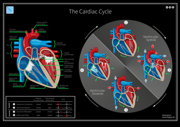
The cycle diagram depicts one heartbeat of the continuously repeating
cardiac cycle
, namely:
ventricular diastole
followed by
ventricular systole
, etc.—while coordinating with
atrial systole
followed by
atrial diastole
, etc. The cycle also correlates to key
electrocardiogram
tracings: the
T wave
(which indicates ventricular diastole); the
P wave
(atrial systole); and the
QRS
'spikes' complex (ventricular systole)—all shown as color purple-in-black segments.
[
1
]
[
2
]
The Cardiac Cycle: Valve Positions, Blood Flow, and ECG
The parts of a
QRS complex
and adjacent deflections. Re the cardiac cycle,
atrial systole
begins at the P wave;
ventricular systole
begins at the Q deflection of the QRS complex.
A
Wiggers diagram
illustrate events and details of the cardiac cycle with electrographic trace lines, which depict (vertical) changes in a parameter's value as time elapses left-to-right.
[
2
]
The ventricular "diastole", or relaxation, begins with "isovolumic relaxation", then proceeds through three sub-stages of inflow, namely: "rapid inflow", "diastasis", and "atrial systole". During the "diastole" period, the "ventricular volume" increases (see red-line tracing), beginning after the vertical bar at
"Aortic valve closes"
and ending with the vertical bar at R in the QRS complex. The ventricular "Systole", or contraction, begins with "isovolumic contraction", i.e., with the vertical bar at
"A -V valve closes"
; it ends with completing the "ejection" stage at the bar at
"aortic valve closes"
. During "ejection" stage, the (red-line) tracing of "ventricular volume" falls to its least amount (see
ejection fraction
) as the ventricles pump blood to the pulmonary arteries and to the aorta.
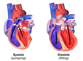
Diastole (at right) normally refers to atria and ventricles at relaxation and expansion together—while refilling with blood returning to the heart. Systole (left) typically refers to
ventricular systole
, during which the ventricles are pumping (or ejecting) blood out of the heart through the aorta and the pulmonary veins.
CGI-animated
graphic of the human heart, sectioned, with motions and timing synced with the Wiggers diagram. The section shows: 1) the opened ventricles contracting once per heartbeat—that is, once per each cardiac cycle; 2) the (partly obscured) mitral valve of the left heart; 3) the tricuspid and pulmonary valves of the right heart—note these paired valves open and close oppositely. + (The aortic valve of the left heart is located below the pulmonary valve, and is completely obscured.) The (unsectioned) atria are seen above the ventricles.
Cardiac diastole: Both AV valves (
tricuspid
in the right heart (light-blue),
mitral
in the left heart (pink)) are open to enable blood to flow directly into both left and right ventricles, where it is collected for the next contraction.
Cardiac (ventricular) systole: Both AV valves (
tricuspid
in the right heart (light-blue),
mitral
in the left heart (pink)) are closed by back-pressure as the ventricles are contracted and their blood volumes are ejected through the newly-opened
pulmonary valve
(dark-blue arrow) and
aortic valve
(dark-red arrow) into the
pulmonary trunk
and aorta respectively.