Cellular dewetting
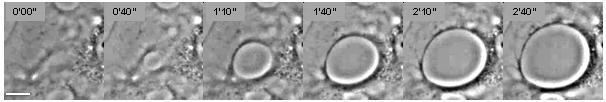
[1] This phenomenon is analogous to the nucleation and growth of dry patches in viscous liquids spreading on a non-wettable substrate (Figure 2).
[2] Cellular dewetting is triggered by several protein toxins from pathogenic bacteria, notably the EDIN-like factors from Staphylococcus aureus and from Clostridium botulinum, as well as edema toxin from Bacillus anthracis.
[3][4] TEMs form in response to the rupture of cytoskeleton physical connections through the cytoplasm due to inhibition of the RhoA/ROCK pathway or to induction of the flux of cyclic-AMP (cAMP) broad signaling molecule.
[6] The driving force responsible for the spontaneous formation of TEM tunnels and their opening is the membrane tension that results from the spreading of cells due to actomyosin relaxation.
This resisting force is referred to as line tension and is uncharacterized at the molecular level.