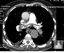
Contrast CT
A small bolus of radio-opaque contrast media is injected into a patient via a peripheral intravenous cannula.
Depending on the vessel being imaged, the volume of contrast is tracked using a region of interest (abbreviated "R.O.I.")
[3] Depending on the purpose of the investigation, there are standardized protocols for time intervals between intravenous radiocontrast administration and image acquisition, in order to visualize the dynamics of contrast enhancements in different organs and tissues.
However, dosages may need to be adjusted or even withheld in patients with risks of iodinated contrast, such as hypersensitivity reactions, contrast-induced nephropathy, effects on thyroid function or adverse drug interactions.
[14] As with CT scans in general, the radiation dose can potentially increase the risk of radiation-induced cancer.