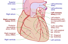
Coronary circulation
Because the rest of the body, and most especially the brain, needs a steady supply of oxygenated blood that is free of all but the slightest interruptions, the heart is required to function continuously.
An anastomosis is an area where vessels unite to form interconnections that normally allow blood to circulate to a region even if there may be partial blockage in another branch.
Therefore, this ability is somewhat restricted in the heart so a coronary artery blockage often results in myocardial infarction causing death of the cells supplied by the particular vessel.
Cardiac veins carry blood with a poor level of oxygen, from the myocardium to the right atrium.
This is because blockage of one coronary artery generally results in death of the heart tissue due to lack of sufficient blood supply from the other branch.
When two arteries or their branches join, the area of the myocardium receives dual blood supply.
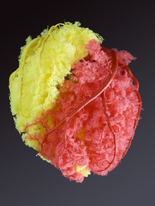
The photograph shows area of heart supplied by the right and the left coronary arteries.
If the papillary muscles are not functioning properly, the mitral valve may leak during contraction of the left ventricle.
The clinical significance of this is that a myocardial infarction involving the PDA is more likely to cause mitral regurgitation.
As a result, most myocardial perfusion occurs during heart relaxation (diastole) when the subendocardial coronary vessels are open and under lower pressure.
Severe ischemia can cause the heart muscle to die from hypoxia, such as during a myocardial infarction.
Chronic moderate ischemia causes contraction of the heart to weaken, known as myocardial hibernation.
[citation needed] In addition to metabolism, the coronary circulation possesses unique pharmacologic characteristics.
When the arteries are healthy, they are capable of autoregulating themselves to maintain the coronary blood flow at levels appropriate to the needs of the heart muscle.
The relatively narrow coronary arteries are commonly affected by atherosclerosis and can become blocked, causing angina or a heart attack.
[citation needed] This article incorporates text from the CC BY book: OpenStax College, Anatomy & Physiology.