Molecular-weight size marker
Protein, DNA, and RNA markers with pre-determined fragment sizes and concentrations are commercially available.
[1] Depending on the running conditions of gel electrophoresis, fragments may have been compressed, disrupting clarity.
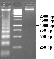
This technique took advantage of logarithmic spacing, and could be used to identify target bands ranging over a length of 20,000 nucleotides.
[3] On the other hand, the size of the DNA pieces are based on the sites where the restriction enzyme cuts.
[5] More recently, another method for constructing DNA molecular-weight size markers is being employed by laboratories.
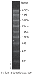
[5] As with experimental samples, the conditions of the gel can affect the molecular-weight size marker that runs alongside them.
The development of a kit including a molecular-weight size marker based on protein fragments began in 1993.
Molecular-weight size markers are most commonly used in SDS-polyacrylamide gel electrophoresis and western blotting.
With all the different types and uses of molecular-weight size markers, it is important to choose the appropriate protein standard.
Besides the most common use, as a way to calculate the molecular weight of the samples, other uses include allowing visual evidence of protein migration and transfer efficiency and are sometimes even used for positive control.
[16] As with DNA electrophoresis, conditions such as buffers, charge/voltage, and concentration should be taken into account when selecting a protein marker.
This technique was improved upon by inventor Eric T. Kool to use circular DNA vectors as a method for producing RNA molecular-weight size markers.
As referred to as the rolling circle method, the improvements of this technique stems from its efficiency in synthesizing RNA oligonucleotides.
From the circular DNA template, single-stranded RNA varying in length from 4-1500 bp can be produced without the need for primers and by recycling nucleotide triphosphate.
In comparison to PCR, the synthetic circle method produces RNA oligonucleotides without the need for polymerase nor a thermal cycler.
Markers are dissolved in a storage buffer, such as EDTA, and can have a shelf life of up to 2 years when stored at -80 °C.
To use the marker, such as for northern blot analysis, it is first thawed, and then stained so that it is detectable on a gel electrophoresis.
However, both RNA and DNA have a negative linear slope between their migration distance and logarithmic molecular weight.
Any glassware that is to come into contact with RNA should be pretreated with diethylpyrocarbonate (DEPC) and plastic materials should be disposable.
Sodium dodecyl sulfate (SDS) interacts with proteins, denaturing them, and giving them a negative charge.
[27] Many kinds of molecular-weight size markers exist, and each possess unique characteristics, lending to their involvement in a number of biological techniques.
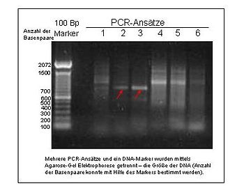
PCR (polymerase chain reaction) is a DNA amplification technique that can be applied to various types of fragments.
These type of markers are based on PCR primers and are categorized as DNA sequence polymorphism.
[28] Although technically speaking, DNA sequence polymorphism has been going on since the use of RFLP in the 1960s, the analysis has changed significantly over the years.