Tooth decay
[12] The earliest sign of a new carious lesion is the appearance of a chalky white spot on the surface of the tooth, indicating an area of demineralization of enamel.
A lesion that appears dark brown and shiny suggests dental caries were once present, but the demineralization process has stopped, leaving a stain.
Once the decay passes through the enamel, the dentinal tubules, which have passages to the nerve of the tooth, become exposed, resulting in pain that can be transient, temporarily worsening with exposure to heat, cold, or sweet foods and drinks.
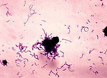
[17][18][19] Four things are required for caries to form: a tooth surface (enamel or dentin), caries-causing bacteria, fermentable carbohydrates (such as sucrose), and time.
These organisms can produce high levels of lactic acid following fermentation of dietary sugars and are resistant to the adverse effects of low pH, properties essential for cariogenic bacteria.
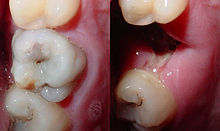
[31] If demineralization continues over time, enough mineral content may be lost so that the soft organic material left behind disintegrates, forming a cavity or hole.
As time progresses, the pH returns to normal due to the buffering capacity of saliva and the dissolved mineral content of tooth surfaces.
In very severe cases where oral hygiene is very poor and where the diet is very rich in fermentable carbohydrates, caries may cause cavities within months of tooth eruption.
[36] Possible contributing factors that have been investigated include systemic factors such as high levels of dioxins or polychlorinated biphenyl (PCB) in the mother's milk, premature birth and oxygen deprivation at birth, and certain disorders during the child's first 3 years such as mumps, diphtheria, scarlet fever, measles, hypoparathyroidism, malnutrition, malabsorption, hypo-vitaminosis D, chronic respiratory diseases, or undiagnosed and untreated coeliac disease, which usually presents with mild or absent gastrointestinal symptoms.
Reduced salivary flow rate is associated with increased caries since the buffering capability of saliva is not present to counterbalance the acidic environment created by certain foods.
Tetrahydrocannabinol (THC), the active chemical substance in cannabis, also causes a nearly complete occlusion of salivation, known in colloquial terms as "cotton mouth".
[69] It is still unknown if the identification of high-risk individuals can lead to more effective long-term patient management that prevents caries initiation and arrests or reverses the progression of lesions.
[75] Tooth enamel is a highly mineralized acellular tissue, and caries act upon it through a chemical process brought on by the acidic environment produced by bacteria.
[78] A slight remineralization of enamel occurs in the dark zone, which serves as an example of how the development of dental caries is an active process with alternating changes.
[89] Early, uncavitated caries is often diagnosed by blowing air across the suspect surface, which removes moisture and changes the optical properties of the unmineralized enamel.
[97] Rampant caries may be seen in individuals with xerostomia, poor oral hygiene, stimulant use (due to drug-induced dry mouth[98]), and/or large sugar intake.
Alternative methods of oral hygiene also exist around the world, such as the use of teeth cleaning twigs such as miswaks in some Middle Eastern and African cultures.
[113] For children, the American Dental Association and the European Academy of Paediatric Dentistry recommend limiting the frequency of consumption of drinks with sugar, and not giving baby bottles to infants during sleep (see earlier discussion).
Sealants can wear out and fail to prevent access of food and plaque bacteria inside pits and fissures and need to be replaced so they must be checked regularly by dental professionals.
American Dental Association Council suggest that for children <3 years old, caregivers should begin brushing their teeth by using fluoridated toothpaste with an amount no more than a smear.
[129] Many dental professionals include application of topical fluoride solutions as part of routine visits and recommend the use of xylitol and amorphous calcium phosphate products.
However, a Cochrane systematic review found no evidence to suggest that taking fluoride systemically daily in pregnant women was effective in preventing dental decay in their offspring.
A Cochrane review of this technique looked at Er:YAG (erbium-doped yttrium aluminium garnet), Er,Cr:YSGG (erbium, chromium: yttrium-scandium-gallium-garnet) and Nd:YAG (neodymium-doped yttrium aluminium garnet) lasers and found that although people treated with lasers (compared to a conventional dental "drill") experienced less pain and had a lesser need for dental anaesthesia, that overall there was little difference in caries removal.
Recent studies from the University of Michigan demonstrated that SDF is effective in stopping tooth decay when applied to the teeth of young children.
[153] The number of cases has decreased in some developed countries, and this decline is usually attributed to increasingly better oral hygiene practices and preventive measures such as fluoride treatment.
Skulls dating from a million years ago through the Neolithic period show signs of caries, including those from the Paleolithic and Mesolithic ages.
[160] Learning their training from apprenticeships, these health providers were quite successful in ending tooth pain and likely prevented systemic spread of infections in many cases.
[169] Pierre Fauchard, known as the father of modern dentistry, was one of the first to reject the idea that worms caused tooth decay and noted that sugar was detrimental to the teeth and gingiva.
[174] The prevalence of caries increased dramatically in the 19th century, as the Industrial Revolution made certain items, such as refined sugar and flour, readily available.
[160] The diet of the "newly industrialized English working class"[160] then became centered on bread, jam, and sweetened tea, greatly increasing both sugar consumption and caries.


|
No data
<50
50–60
60–70
70–80
80–90
90–100
|
100–115
115–130
130–138
138–140
140–142
>142
|
