Dental fluorosis
[3][needs update] Dental fluorosis appears as a range of visual changes in enamel[4] causing degrees of intrinsic tooth discoloration, and, in some cases, physical damage to the teeth.
[1] People with fluorosis are relatively resistant to dental caries (tooth decay caused by bacteria),[2] although there may be cosmetic concern.
[6] There are individual variations in clinical fluorosis manifestation which are highly dependent on the duration, timing, and dosage of fluoride exposure.
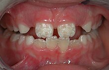
The clinical manifestation of mild dental fluorosis is mostly characterised a snow flaking appearance that lack a clear border, opaque, white spots, narrow white lines following the perikymata or patches as the opacities may coalesce with an intact, hard and smooth enamel surface on most of the teeth.
In dental enamel, fluorosis causes subsurface porosity or hypomineralizations, which extend toward the dentinal-enamel junction as the condition progresses and the affected teeth become more susceptible to staining.
Due to diffusion of exogenous ions (e.g., iron and copper), stains develop into the increasingly and abnormally porous enamel.
[12] It scores the spectrum of fluorotic changes in enamel from 0 to 9, allowing more precise definition of mild and severe cases.
[13] Dental fluorosis is caused by a higher than normal amount of fluoride ingestion whilst teeth are forming.
[17] The severity of dental fluorosis depends on the amount of fluoride exposure, the age of the child, individual response, weight, degree of physical activity, nutrition, and bone growth.
[20] The last of these sources is directly or indirectly responsible for 40% of all fluorosis, but the resulting effect due to water fluoridation is largely and typically aesthetic.
[22] Dental fluorosis has been growing in the United States concurrent with fluoridation of municipal water supplies, although disproportionately by race.
[24] The 2011-12 NHANES figures documented another 31% overall increase among American teens since the previous decade, with a total adolescent population impact of 61% afflicted.
[2][12] Fluoride may also indirectly alter the action of protease via a decrease in the availability of free calcium ions in the mineralization environment.
The condition is more prevalent in rural areas where drinking water is derived from shallow wells or hand pumps.
[citation needed] It is also more likely to occur in areas where the drinking water has a fluoride content greater than 1 ppm (part per million).
[12] In 1931, 3 different groups of scientists around the world published their discoveries that this condition was caused by fluoride in drinking water during childhood.
[37] Through epidemiological studies in the US, Henry Trendley Dean helped to identify a causal link between high concentrations of fluoride in the drinking water and mottled enamel.