Dissection
Prevention of transmission includes the wearing of protective gear, ensuring the environment is clean, dissection technique[2] and pre-dissection tests to specimens for the presence of HIV and hepatitis viruses.
[6] Human dissections were carried out by the Greek physicians Herophilus of Chalcedon and Erasistratus of Chios in the early part of the third century BC.
[10] While there was a deep taboo in Greek culture concerning human dissection, there was at the time a strong push by the Ptolemaic government to build Alexandria into a hub of scientific study.
[15] The images showing how to kill most effectively depending on the game being hunted relay an intimate knowledge of both external and internal anatomy as well as the relative importance of organs.
[15] Early in the history of India (2nd to 3rd century), the Arthashastra described the 4 ways that death can occur and their symptoms: drowning, hanging, strangling, or asphyxiation.
The process involved the loosening of the tissues in streams of water before the outer layers were sloughed off with soft implements to reach the musculature.
[17] In Saudi Arabia, whose law is completely dictated by Shari'ah, autopsy is viewed poorly by the population but can be compelled in criminal cases;[17] human dissection is sometimes found at university level.
[17] Human dissection is present in the modern day Islamic world, but is rarely published on due to the religious and social stigma.
Tibetans had adopted the practice of sky burial because of the country's hard ground, frozen for most of the year, and the lack of wood for cremation.
[25][26] Throughout the history of Christian Europe, the dissection of human cadavers for medical education has experienced various cycles of legalization and proscription in different countries.
[8][34] Frederick II (1194–1250), the Holy Roman Emperor, decreed that any that were studying to be a physician or a surgeon must attend a human dissection, which would be held no less than every five years.
[10] At this time, autopsies were carried out by a team consisting of a Lector, who lectured; the Sector, who did the dissection; and the Ostensor, who pointed to features of interest.
[10][36] The Catholic Church is known to have ordered an autopsy on conjoined twins Joana and Melchiora Ballestero in Hispaniola in 1533 to determine whether they shared a soul.
Though most chose to focus on the external surfaces of the body, some like Michelangelo Buonarotti, Antonio del Pollaiuolo, Baccio Bandinelli, and Leonardo da Vinci sought a deeper understanding.
However, there were no provisions for artists to obtain cadavers, so they had to resort to unauthorised means, as indeed anatomists sometimes did, such as grave robbing, body snatching, and murder.
[38] In modern Europe, dissection is routinely practised in biological research and education, in medical schools, and to determine the cause of death in autopsy.
The permission was quite limited: by the mid-18th century, the Royal College of Physicians and Company of Barber-Surgeons were the only two groups permitted to carry out dissections, and had an annual quota of ten cadavers between them.
[41] By the 21st century, the availability of interactive computer programs and changing public sentiment led to renewed debate on the use of cadavers in medical education.
The Peninsula College of Medicine and Dentistry in the UK, founded in 2000, became the first modern medical school to carry out its anatomy education without dissection.
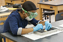
[42] In the United States, dissection of frogs became common in college biology classes from the 1920s, and were gradually introduced at earlier stages of education.
Other popular animals for high-school dissection at the time of that survey were, among vertebrates, fetal pigs, perch, and cats; and among invertebrates, earthworms, grasshoppers, crayfish, and starfish.
Other states including Arizona, Hawaii, Minnesota, Texas, and Utah have more general policies on opting out on moral, religious, or ethical grounds.
[56] Studies show that some students reluctantly participate in animal dissection out of fear of real or perceived punishment or ostracism from their teachers and peers, and many do not speak up about their ethical objections.
At Stanford Medical School, software combines X-ray, ultrasound and MRI imaging for display on a screen as large as a body on a table.
Those in favor of dissection alternatives point to studies which have shown that computer-based teaching methods "saved academic and nonacademic staff time ... were considered to be less expensive and an effective and enjoyable mode of student learning [and] ... contributed to a significant reduction in animal use" because there is no set-up or clean-up time, no obligatory safety lessons, and no monitoring of misbehavior with animal cadavers, scissors, and scalpels.
Some programs also allow educators to customize lessons and include built-in test and quiz modules that can track student performance.
Furthermore, animals (whether dead or alive) can be used only once, while non-animal resources can be used for many years—an added benefit that could result in significant cost savings for teachers, school districts, and state educational systems.
[61] Several peer-reviewed comparative studies examining information retention and performance of students who dissected animals and those who used an alternative instruction method have concluded that the educational outcomes of students who are taught basic and advanced biomedical concepts and skills using non-animal methods are equivalent or superior to those of their peers who use animal-based laboratories such as animal dissection.
[64][65] Some reports state that students' confidence, satisfaction, and ability to retrieve and communicate information was much higher for those who participated in alternative activities compared to dissection.
Three separate studies at universities across the United States found that students who modeled body systems out of clay were significantly better at identifying the constituent parts of human anatomy than their classmates who performed animal dissection.