Cell division
For simple unicellular microorganisms such as the amoeba, one cell division is equivalent to reproduction – an entire new organism is created.
On a larger scale, mitotic cell division can create progeny from multicellular organisms, such as plants that grow from cuttings.
[5][6] After growth from the zygote to the adult, cell division by mitosis allows for continual construction and repair of the organism.
[9] A great deal of cellular infrastructure is involved in ensuring consistency of genomic information among generations.
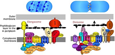
A tubulin-like protein, FtsZ plays a critical role in formation of a contractile ring for the cell division.
The amitotic or mitotic cell divisions are more atypical and diverse among the various groups of organisms, such as protists (namely diatoms, dinoflagellates, etc.)
[citation needed] In meiosis I, the homologous chromosomes are paired before being separated and distributed between two daughter cells.
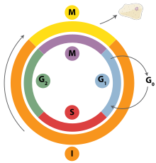
[18] During G2, the cell undergoes the final stages of growth before it enters the M phase, where spindles are synthesized.
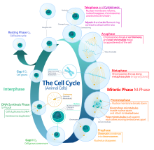
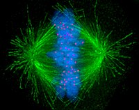
The nuclear envelope begins to be broken down in this stage, long strands of chromatin condense to form shorter more visible strands called chromosomes, the nucleolus disappears, and the mitotic spindle begins to assemble from the two centrosomes.
[22] This process is evidenced to be caused in a large part by the highly conserved Spo11 protein through a mechanism similar to that seen with topoisomerase in DNA replication and transcription.
This breakdown then allows the spindle apparatus growing from the centrosome to attach to the kinetochores on the sister chromatids.
Stable attachment of the spindle apparatus to the kinetochores on the sister chromatids will ensure error-free chromosome segregation during anaphase.
[27] Anaphase is a very short stage of the cell cycle and it occurs after the chromosomes align at the mitotic plate.
[26] This abrupt shift is caused by the activation of the anaphase-promoting complex and its function of tagging degradation of proteins important toward the metaphase-anaphase transition.
One of these proteins that is broken down is securin which through its breakdown releases the enzyme separase that cleaves the cohesin rings holding together the sister chromatids thereby leading to the chromosomes separating.
[30] Additionally, in this phase, the activation of the anaphase promoting complex through the association with Cdh-1 begins the degradation of mitotic cyclins.
This as a result leads to cytokinesis producing unequal daughter cells containing completely different amounts or concentrations of fate-determining molecules.
[41] These checkpoint kinases phosphorylate p53, which stimulates the production of different enzymes associated with DNA repair.
These cyclin-cdk complexes phosphorylate the Retinoblastoma (Rb) protein, a tumor suppressor bound with the E2F family of transcription factors.
[43] If DNA is damaged, the cell can also alter the Akt pathway in which BAD is phosphorylated and dissociated from Bcl2, thus inhibiting apoptosis.
If this pathway is altered by a loss of function mutation in Akt or Bcl2, then the cell with damaged DNA will be forced to undergo apoptosis.
PUMA is a pro-apoptotic protein that rapidly induces apoptosis by inhibiting the anti-apoptotic Bcl-2 family members.
[48] At the beginning of the 19th century, various hypotheses circulated about cell proliferation, which became observable in plant and animal organisms as a result of advances in microscopy.
The Belgian botanist Barthélemy Charles Joseph Dumortier must be regarded as the first discoverer of cell division.
In 1832, he described cell division in simple aquatic plants (French 'conferve') as follows (translated from French to English): "The development of the conferve is as simple as its structure; it takes place by the attachment of new cells to the old, and this attachment always takes place from the end.
It is impossible to determine this, but it is always true that it later appears double when united, and that when two cells naturally separate, each of them is closed at both ends.
"[53] In 1835, the German botanist and physician Hugo von Mohl described plant cell division in much greater detail in his dissertation on freshwater and seawater algae for his PhD thesis in medicine and surgery:[54] "Among the most obscure phenomena of plant life is the manner in which the newly developing cells are formed.
[55] The German-Polish physician Robert Remak suspected that he had already discovered animal cell division in the blood of chicken embryos in 1841,[56] but it was not until 1852 that he was able to confirm animal cell division for the first time in bird embryos, frog larvae and mammals.