Doppler optical coherence tomography
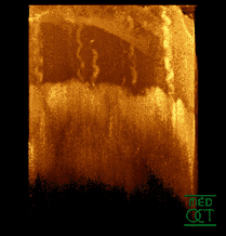
Optical coherence tomography (OCT) is a technique that displays images of the tissue by using the backscattered light.
[citation needed] Not only conserving the excellence of OCT, doppler optical coherence tomography also combines the doppler effect principle as a whole, which result in tomographic images with high resolution.
[1] Not only conserving the excellence of OCT, Doppler Optical Coherence Tomography also combines the Doppler effect principle as a whole, which results in tomographic images with high resolution with static and moving constituents.
The basic principle of Phase-resolved Doppler OCT uses the phase change between sequential A-line scans for velocity image reconstruction.
[1] This improvement shows prominent increase in the scanning speed and sensitivity, which makes it possible to image in vivo tissue microcirculation in human skin.
[4] Because of its exceptionally high spatial resolution and velocity sensitivity, Doppler OCT has its own position in the field of biomedical research and clinical medicine.
[5] Doppler Optical Coherence Tomography is an extension of OCT, where it combines the Doppler effect principle to achieve high resolution tomographic images in biological tissues.
And because of its high resolution and velocity sensitivity, there are many applications in the medical field.
are wave vectors of incident and out scattered light, and v is the velocity of the moving particle the instrument is detecting.
Doppler OCT measures the light backscattered from the sample medium.
Defining the angle between the particle flow and the incident light beam is θ, the Doppler shift is then simplified to where
It also uses a fiber optic Michelson interferometer with a broadband light as a source.
Time domain Doppler OCT uses the spectrogram method to create the image.
[1] The short-time fast Fourier transformation (STFFT) is used to calculate the power spectrum.
is the centroid frequency and θ is the angle between ki and v.[1] When dealing with high speed imaging, due to many factors, the velocity sensitivity is unsatisfied.
When the difference in flow velocity becomes larger, the Doppler frequency spectrum becomes wider.
[1] There are two methods: a spectrometer based system[6][7] and a swept laser source based system [8][9][10][11][12] to obtain high velocity sensitivity, high imaging speed, and various velocity range [13][14][15][16][17]