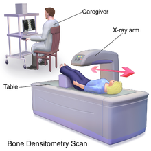
Dual-energy X-ray absorptiometry
Dual-energy X-ray absorptiometry (DXA, or DEXA[1]) is a means of measuring bone mineral density (BMD) with spectral imaging.
One type of DXA scanner uses a cerium filter with a tube voltage of 80 kV, resulting in effective photon energies of about 40 and 70 keV.
[2] Another type of DXA scanner uses a samarium filter with a tube voltage of 100 kV, which produces effective energies of 47 and 80 keV.
To avoid an overestimation of bone mineral deficits, BMD scores are commonly compared to reference data for the same gender and age (by calculating a Z-score).
Despite DXA technology's problems with estimating volume, it is still a fairly accurate measure of bone mineral content.
Methods to correct for this shortcoming include the calculation of a volume that is approximated from the projected area measure by DXA.
The ISCD states that there is no clearly understood correlation between BMD and the risk of a child's sustaining a fracture; the diagnosis of osteoporosis in children cannot be made on the basis of densitometry criteria.
[9] Some clinics may routinely carry out DXA scans on pediatric patients with conditions such as nutritional rickets, lupus, and Turner syndrome.
[10] DXA has been demonstrated to measure skeletal maturity[11] and body fat composition[12] and has been used to evaluate the effects of pharmaceutical therapy.
[13] It may also aid pediatricians in diagnosing and monitoring treatment of disorders of bone mass acquisition in childhood.
[16][17] DXA scans can also be used to measure total body composition and fat content with a high degree of accuracy comparable to hydrostatic weighing with a few important caveats.
[19] It has been suggested that, while very accurately measuring minerals and lean soft tissue (LST), DXA may provide skewed results due to its method of indirectly calculating fat mass by subtracting it from the LST and/or body cell mass (BCM) that DXA actually measures.
[20] DXA scans have been suggested as useful tools to diagnose conditions with an abnormal fat distribution, such as familial partial lipodystrophy.
The radiation dose of current DEXA systems is small,[24] as low as 0.001 mSv, much less than a standard chest or dental x-ray.
For example, in Victoria, an individual performing DXA scans is required to completed a recognised course in safe use of bone mineral densitometers.