Trisomy 18
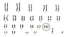
Trisomy 18, also known as Edwards syndrome, is a genetic disorder caused by the presence of a third copy of all or part of chromosome 18.
Most cases of trisomy 18 occur due to problems during the formation of the reproductive cells or during early development.
[3] Occasionally, not all cells have the extra chromosome, known as mosaic trisomy, and symptoms in these cases may be less severe.
[5] Children born with Edwards' syndrome may have some or all of these characteristics: kidney malformations, structural heart defects at birth (e.g., ventricular septal defect, atrial septal defect, patent ductus arteriosus), intestines protruding outside the body (omphalocele), esophageal atresia, intellectual disability, developmental delays, growth deficiency, feeding difficulties, breathing difficulties, and arthrogryposis (a muscle disorder that causes multiple joint contractures at birth).
[7][8] Some physical malformations associated with Edwards' syndrome include small head (microcephaly) accompanied by a prominent back portion of the head (occiput), low-set, malformed ears, abnormally small jaw (micrognathia), cleft lip/cleft palate, upturned nose, narrow eyelid openings (blepharophimosis), widely spaced eyes (ocular hypertelorism), drooping of the upper eyelids (ptosis), a short breast bone, clenched hands, choroid plexus cysts, underdeveloped thumbs and/or nails, absent radius, webbing of the second and third toes, clubfoot or rocker bottom feet, and in males, undescended testicles.
The most common intracranial anomaly is the presence of choroid plexus cysts, which are pockets of fluid on the brain.
[7] Although uncommon in the syndrome, trisomy 18 causes a large portion of prenatally diagnosed cases of Dandy–Walker malformation.
Numerical errors can arise at either of the two meiotic divisions and cause the failure of a chromosome to segregate into the daughter cells (nondisjunction).
(Although an embryo could inherit a trisomy from both parents, it is, as a rule, extremely rare, and worse in terms of clinical perspective and prognosis.)
[2] Levels of PAPP-A, AFP, and uE3 are generally decreased during pregnancy, and free beta HCG is elevated.
[21] Though rare, some persons with Trisomy 18 survive into their twenties and thirties with the current eldest being well over 50 years.
Current ongoing research at the University of Michigan shows survival rate with full interventions is about 90% until the first birthday, and 80% until 5 years.