Arthropod exoskeleton
Typically the mineral crystals, mainly calcium carbonate, are deposited among the chitin and protein molecules in a process called biomineralization.
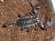
The dorsal tergum, ventral sternum, and the lateral pleura form the hardened plates or sclerites of a typical body segment.
In either case, in contrast to the carapace of a tortoise or the cranium of a vertebrate, the exoskeleton has little ability to grow or change its form once it has matured.
A typical arthropod exoskeleton is a multi-layered structure with four functional regions: epicuticle, procuticle, epidermis and basement membrane.
The strength of the exoskeleton is provided by the underlying procuticle, which is in turn secreted by the epithelial cells in the epidermis,[2] which begins as a tough, flexible layer of chitin.
By varying the types of interaction between the proteins and chitins, the insect metabolism produces regions of exoskeleton that differ in their wet and dry behaviour, their colour and their mechanical properties.
[7] The inner layer is not as highly sclerotised, and is correspondingly softer but tougher; it resists tensile stresses but is liable to failure under compression.
Sclerites may be simple protective armour, but also may form mechanical components of the exoskeleton, such as in the legs, joints, fins or wings.
However, in most Arthropoda the bodily tagmata are so connected and jointed with flexible cuticle and muscles that they have at least some freedom of movement, and many such animals, such as the Chilopoda or the larvae of mosquitoes are very mobile indeed.
The internal surface of the exoskeleton is often infolded, forming a set of structures called apodemes that serve for the attachment of muscles, and functionally amounting to endoskeletal components.
[citation needed] Within entomology, the term glabrous is used to refer to those parts of an insect's body lacking in setae (bristles) or scales.
In some groups of animals, most conspicuously the Crustacea, the matrix is greatly enriched with, or even dominated by, hard minerals, usually calcite or similar carbonates that form much of the exoskeleton.
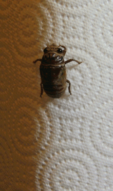
[11] The difference between the unmodified and modified forms of chitinous arthropodan exoskeletons can be seen by comparing the body wall of say a bee larva, in which modification is minimal, to any armoured species of beetle, or the fangs of a spider.
The chemical and physical nature of the arthropod exoskeleton limits its ability to stretch or change shape as the animal grows.
Secondly, often a major injury in one phase, such as the loss of a leg from an insect nymph, or a claw from a young crab, can be repaired after one or two stages of ecdysis.
Similarly, delicate parts that need periodic replacement, such as the outer surfaces of the eye lenses of spiders, or the urticating hairs of caterpillars, can be shed, making way for new structures.