Functional magnetic resonance imaging
[4] Since the early 1990s, fMRI has come to dominate brain mapping research because it does not involve the use of injections, surgery, the ingestion of substances, or exposure to ionizing radiation.
[11] During the late 19th century, Angelo Mosso invented the 'human circulation balance', which could non-invasively measure the redistribution of blood during emotional and intellectual activity.
[13] Angelo Mosso investigated several critical variables that are still relevant in modern neuroimaging such as the 'signal-to-noise ratio', the appropriate choice of the experimental paradigm and the need for the simultaneous recording of differing physiological parameters.
[13] Mosso's manuscripts do not provide direct evidence that the balance was really able to measure changes in cerebral blood flow due to cognition,[13] however a modern replication performed by David T Field[14] has now demonstrated—using modern signal processing techniques unavailable to Mosso—that a balance apparatus of this type is able to detect changes in cerebral blood volume related to cognition.
In a seminal 1990 study based on earlier work by Thulborn et al., Ogawa and colleagues scanned rodents in a strong magnetic field (7.0 T) MRI.
[16] The first attempt to detect the regional brain activity using MRI was performed by Belliveau and colleagues[17] at Harvard University using the contrast agent Magnevist, a paramagnetic substance remaining in the bloodstream after intravenous injection.
Kenneth Kwong and colleagues, using both gradient-echo and inversion recovery echo-planar imaging (EPI) sequence at a magnetic field strength of 1.5 T published studies showing clear activation of the human visual cortex.
Ogawa and Ugurbil conducted a similar study using a higher magnetic field (4.0 T) in Ugurbil's laboratory at the University of Minnesota, generating higher resolution images that showed activity largely following the gray matter of the brain, as would be expected; in addition, they showed that fMRI signal depended on a decrease in T2*, consistent with the BOLD mechanism.
Bandettini and colleagues used EPI at 1.5 T to show activation in the primary motor cortex, a brain area at the last stage of the circuitry controlling voluntary movements.
Initial results show there is more inflow than consumption of glucose in regions such as the amygdala, basal ganglia, thalamus and cingulate cortex, all of which are recruited for fast responses.
Experimental paradigms such as staggering when a stimulus is presented at various trials can improve temporal resolution, but reduces the number of effective data points obtained.
Neuronal activity related to the act of seeing lasts for more than 100 ms. A fast reaction, such as swerving to avoid a car crash, takes around 200 ms. By about half a second, awareness and reflection of the incident sets in.
Activation locations detected by BOLD fMRI in cortical areas (brain surface regions) are known to tally with CBF-based functional maps from PET scans.
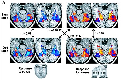
Nearby regions such as the pulvinar nucleus were not stimulated for this task, indicating millimeter resolution for the spatial extent of the BOLD response, at least in thalamic nuclei.
[38] The BOLD response can also be affected by a variety of factors, including disease, sedation, anxiety, medications that dilate blood vessels,[39] and attention (neuromodulation)[40].
The scanning process acquires the MR signal in k-space, in which overlapping spatial frequencies (that is repeated edges in the sample's volume) are each represented with lines.
[57] Physiological noise is from head and brain movement in the scanner from breathing, heart beats, or the subject fidgeting, tensing, or making physical responses such as button presses.
Even when whole-brain analysis is done, to interpret the final results, that is to figure out which regions the active voxels fall in, one has to align the functional image to the structural one.
While this is conceptually similar to motion correction, the changes required are more complex than just translation and rotation, and hence optimization even more likely to depend on the first transformations in the chain that is checked.
[77][78] Both block and event-related designs are based on the subtraction paradigm, which assumes that specific cognitive processes can be added selectively in different conditions.
[74][clarification needed] Overlapping signals in fMRI are a significant challenge in cognitive neuroscience research, particularly when multiple stimuli or tasks are presented in close temporal proximity.
This causes signals from different brain processes to overlap, making it difficult to differentiate which neural activity is associated with specific stimuli or tasks.
Advances in analytical methods, such as specialized tools for optimizing experimental designs, are crucial for mitigating the effects of signal overlap and improving the reliability of fMRI studies.
Neuroimaging methods such as fMRI and MRI offer a measure of the activation of certain brain areas in response to cognitive tasks engaged in during the scanning process.
Forward inference supports the dual process theory by demonstrating that there are two qualitatively different brain activation patterns when distinguishing between "remember vs. know judgments".
[92] Scanning sessions also subject participants to loud high-pitched noises from Lorentz forces induced in the gradient coils by the rapidly switching current in the powerful static field.
[95][96] However, genotoxic (i.e., potentially carcinogenic) effects of MRI scanning have been demonstrated in vivo and in vitro,[97][98][99][100] leading a recent review to recommend "a need for further studies and prudent use in order to avoid unnecessary examinations, according to the precautionary principle".
Other biomarkers now looked at to provide better contrast include temperature, acidity/alkalinity (pH), calcium-sensitive agents, neuronal magnetic field, and the Lorentz effect.
[103] Other studies have shown the brain activity that characterizes men's preference for sports cars, and even differences between Democrats and Republicans in their reaction to campaign commercials with images of the 9/11 attacks.
These companies depend on evidence such as that from a study by Joshua Greene at Harvard University suggesting the prefrontal cortex is more active in those contemplating lying.