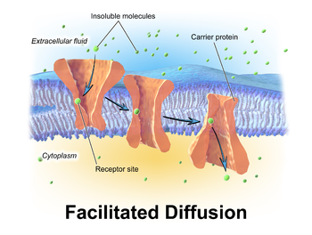
Facilitated diffusion
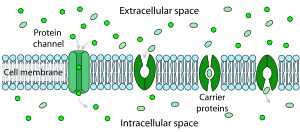
These channels are gated, meaning that they open and close, and thus deregulate the flow of ions or small polar molecules across membranes, sometimes against the osmotic gradient.
They are transported through aqueous compartments of cells or through extracellular space by water-soluble carriers (e.g. retinol binding protein).
Their transport must therefore be "facilitated" by proteins that span the membrane and provide an alternative route or bypass mechanism.
Facilitated diffusion is the main mechanism behind the binding of Transcription Factors (TFs) to designated target sites on the DNA molecule.
Bauer & Metzler (2013)[4] therefore carried out an experiment using a bacterial genome in which they investigated the average time for TF – DNA binding to occur.
[5] In prokaryotic bacteria cells such as E. coli, facilitated diffusion is required in order for regulatory proteins to locate and bind to target sites on DNA base pairs.
The in vivo model mentioned above clearly explains 3-D and 1-D diffusion along the DNA strand and the binding of proteins to target sites on the chain.
[7] The oxygen affinity with hemoglobin on red blood cell surfaces enhances this bonding ability.
[9] This mechanism of facilitated diffusion of oxygen by hemoglobin or myoglobin was discovered and initiated by Wittenberg and Scholander.
Facilitated diffusion helps in the release of accumulated glucose into the extracellular space adjacent to the blood capillary.