Febrile neutrophilic dermatosis
[3][4][5] Acute, tender, erythematous plaques, nodes, pseudovesicles and, occasionally, blisters with an annular or arciform pattern occur on the head, neck, legs, and arms, particularly the back of the hands and fingers.
An underlying condition (streptococcal infection, inflammatory bowel disease, nonlymphocytic leukemia and other hematologic malignancies, solid tumors, pregnancy) is found in up to 50% of cases.
Attacks of SS may precede the hematologic diagnosis by 3 months to 6 years, so that close evaluation of patients in the “idiopathic” group is required.
[7] It is known that treatment with corticosteroids often leads to favorable outcomes, and the frequencies of human leukocyte antigen (HLA) types B54 and Cw1 are notably high, indicating their association with the condition.
[8] The clinical differential diagnosis includes pyoderma gangrenosum, infection, erythema multiforme, adverse drug reactions, and urticaria.
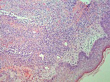
Skin biopsy shows a papillary and mid-dermal mixed infiltrate of polymorphonuclear leukocytes with nuclear fragmentation and histiocytic cells.
The infiltrate is predominantly perivascular with endothelial-cell swelling in some vessels, but vasculitic changes (blood clots; deposition of fibrin, complement, or immunoglobulins within the vessel walls; red blood cell extravasation;inflammatory infiltration of vascular walls) are absent in early lesions.Perivasculitis occurs secondarily, because of cytokines released by the lesional neutrophils.
[citation needed] Sweet described a disease with four features: fever; leukocytosis; acute, tender, red plaques; and a papillary dermal infiltrate of neutrophils.
Oral potassium iodide or colchicine may induce rapid resolution.Patients who have a potential systemic infection or in whom corticosteroids are contraindicated can use these agents as a first-line therapy.
Seventeen of 18 patients had a good initial response; fever and arthralgias were markedly attenuated within 48 hours, and eruptions cleared between 7 and 14 days.