Fluorodeoxyglucose (18F)
[2] The subsequent discovery in 1980 that [18F]FDG accumulates in tumors underpins the evolution of PET as a major clinical tool in cancer diagnosis.
In 1968, Dr. Josef Pacák, Zdeněk Točík and Miloslav Černý at the Department of Organic Chemistry, Charles University, Czechoslovakia were the first to describe the synthesis of FDG.
Brain images obtained with an ordinary (non-PET) nuclear scanner demonstrated the concentration of [18F]FDG in that organ (see history reference below).
[8] To achieve this chemistry, the [18F]F− is separated from the aqueous solvent by trapping it on an ion-exchange column, and eluted with an acetonitrile solution of 2,2,2-cryptand and potassium carbonate.
After picking up a proton H+ from a hydronium ion in its aqueous environment, the molecule becomes glucose-6-phosphate labeled with harmless nonradioactive "heavy oxygen" in the hydroxyl at the C-2 position.
In practice, patients who have been injected with [18F]FDG are told to avoid the close vicinity of especially radiation-sensitive persons, such as infants, children and pregnant women, for at least 12 hours (7 half-lives, or decay to 1⁄128 the initial radioactive dose).
[13] IBA Molecular North America and Zevacor Molecular, both of which are owned by Illinois Health and Science (IBAM having been purchased as of 1 August 2015), Siemens' PETNET Solutions (a subsidiary of Siemens Healthcare), and Cardinal Health[14] are producers in the U.S.[15][16][17][18] The labeled [18F]FDG compound has a relatively short shelf life which is dominated by the physical decay of fluorine-18 with a half-life of 109.8 minutes, or slightly less than two hours.
Still, this half life is sufficiently long to allow shipping the compound to remote PET scanning facilities, in contrast to other medical radioisotopes like carbon-11.
Transport by air allows expanding the distribution area around a [18F]FDG production site to deliver the compound to PET scanning centres even hundreds of miles away.
[citation needed] Recently, on-site cyclotrons with integral shielding and portable chemistry stations for making [18F]FDG have accompanied PET scanners to remote hospitals.
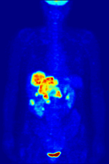
[18F]FDG is taken up by cells, and subsequently phosphorylated by hexokinase (whose mitochondrial form is greatly elevated in rapidly growing malignant tumours).
The patient must then wait about an hour for the sugar to distribute and be taken up into organs which use glucose – a time during which physical activity must be kept to a minimum, in order to minimize uptake of the radioactive sugar into muscles (this causes unwanted artifacts in the scan, interfering with reading especially when the organs of interest are inside the body vs. inside the skull).
Then, the patient is placed in the PET scanner for a series of one or more scans which may take from 20 minutes to as long as an hour (often, only about one-quarter of the body length may be imaged at a time).