Scintigraphy
In contrast, SPECT and positron emission tomography (PET) form 3-dimensional images and are therefore classified as separate techniques from scintigraphy, although they also use gamma cameras to detect internal radiation.
This pulse (scintillation) is usually detected and amplified by a photomultiplier or charge-coupled device elements, and its resulting electrical waveform is processed by computers to provide two- and three-dimensional images of a subject or region of interest.
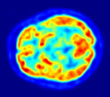
The subjects are injected with special radionuclides which irradiate in the gamma range inside the region of interest, such as the heart or the brain.
Another medical scintillography technique, the Positron-emission tomography (PET), which uses the scintillations provoked by electron-positron annihilation phenomena.
Scintigraphy of the biliary system is called cholescintigraphy and is done to diagnose obstruction of the bile ducts by a gallstone (cholelithiasis), a tumor, or another cause.
[2] The most common indication for lung scintigraphy is to diagnose pulmonary embolism, e.g. with a ventilation/perfusion scan and may be appropriate for excluding PE in pregnancy.
[4] In the ventilation phase of a ventilation/perfusion scan, a gaseous radionuclide xenon or technetium DTPA in an aerosol form (or ideally using Technegas, a radioaerosol invented in Australia by Dr Bill Burch and Dr Richard Fawdry) is inhaled by the patient through a mouthpiece or mask that covers the nose and mouth.
Scintigraphic scanning was invented and proven by Neurologist and Radiologist professor Bernard George Ziedses des Plantes.
In 1970, the Physikalisch-Medizinische Gesellschaft für Neuroradiologie (The Physics and Medical Society for Neuroradiology) instituted the ‘Ziedses des Plantes Medal'.