Fluorescence imaging
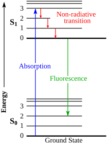
Fluorescence itself, is a form of luminescence that results from matter emitting light of a certain wavelength after absorbing electromagnetic radiation.
The subsequent return to ground state results in emission of fluorescent light that can be detected and measured.
Typically a large scanning device or CCD is used here to measure the intensity and digitally photograph an image.
[1] Since some wavelengths of fluorescence are beyond the range of the human eye, charged-coupled devices (CCD) are used to accurately detect light and image the emission.
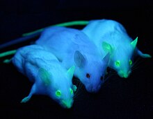
Developing more effective fluorescent proteins is a task that many scientists have taken up in order to improve imaging probe capabilities.
For example, by mutating the F64L gene in jellyfish GFP, the protein is able to more efficiently fluoresce at 37 °C, an important attribute to have when growing cultures in a laboratory.
Mechanisms that have been well described but not necessarily incorporated into practical applications hold promising potential for fluorescence imaging.
Fluorescence resonance energy transfer (FRET) is an extremely sensitive mechanism that produce signaling molecules in the range of 1–10 nm.
[8] Development of more sensitive probes and analytical techniques for laser induced fluorescence can allow for more accurate, up-to-date experimental data.