Photoactivated localization microscopy
[4] The development of PALM as a targeted biophysical imaging method was largely prompted by the discovery of new species and the engineering of mutants of fluorescent proteins displaying a controllable photochromism, such as photo-activatible GFP.
However, the concomitant development of STORM, sharing the same fundamental principle, originally made use of paired cyanine dyes.
A growing number of dyes are used for PALM, STORM and related techniques, both organic fluorophores and fluorescent proteins.
However, if the emission from the two neighboring fluorescent molecules is made distinguishable, i.e. the photons coming from each of the two can be identified, then it is possible to overcome the diffraction limit.
[9] Once a set of photons from a specific molecule is collected, it forms a diffraction-limited spot in the image plane of the microscope.
The error that is made in localizing the center of a point emitter scales to a first approximation as the inverse square root of the number of emitted photons, and if enough photons are collected it is easy to obtain a localization error much smaller than the original point spread function.
The two steps of identification and localization of individual fluorescent molecules in a dense environment where many are present are at the basis of PALM, STORM and their development.
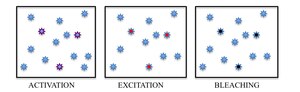
By turning on stochastically sparse subsets of fluorophores with light of a specific wavelength, individual molecules can then be excited and imaged according to their spectra.
To avoid the accumulation of active fluorophores in the sample, which would eventually degrade back to a diffraction-limited image, the spontaneously occurring phenomenon of photobleaching is exploited in PALM, whereas reversible switching between a fluorescent on-state and a dark off-state of a dye is exploited in STORM.
In summary, PALM and STORM are based on collecting under a fluorescent microscope a large number of images each containing just a few active isolated fluorophores.
During each cycle, the density of activated molecules is kept low enough that the molecular images of individual fluorophores do not typically overlap.
In each image of the sequence, the position of a fluorophore is calculated with a precision typically greater than the diffraction limit - in the typical range of a few to tens of nm - and the resulting information of the position of the centers of all the localized molecules is used to build up the super-resolution PALM or STORM image.
[10] The requirement of localizing at the same time multiple fluorophores simultaneously over an extended area determines the reason why these methods are wide-field, employing as a detector a CCD, EMCCD or a CMOS camera.
To determine the axial position of a single fluorophore in the sample the following approaches are currently being used: modification of the point spread function to introduce z-dependent features in the 2D (x,y) image (the most common approach is to introduce astigmatism in the PSF); multiplane detection, where the axial position is determined by comparing two images of the same PSF defocused one with respect to the other; interferometric determination of the axial position of the emitter using two opposed objectives and multiple detectors;[7] use of temporal focusing to confine the excitation/activation; use of light sheet excitation/activation to confine to a few hundred nanometers thick layer arbitrarily positioned along the z-plane within the sample.
The requirement for multiple cycles of activation, excitation and de-activation/bleaching would typically imply extended periods of time to form a PALM/STORM image, and therefore operation on a fixed sample.
The ability to perform live super-resolution imaging using these techniques ultimately depends on the technical limitations of collecting enough photons from a single emitter in a very short time.
[21] These studies have found that, in addition to the standard uncertainty of localization due to the point spread function fitting, self-interference with light scattered by nanoparticles can lead to distortions or displacements of the imaged point spread functions,[20][21] complicating the analysis of such measurements.
These may be possible to limit, however, for example by incorporating metasurface masks which control the angular distribution of light permitted into the measurement system.
[22] PALM and STORM share a common fundamental principle, and numerous developments have tended to make the two techniques even more intertwined.
On the technical side, PALM is performed on a biological specimen using fluorophores expressed exogenously in the form of genetic fusion constructs to a photoactivatable fluorescent protein.
In STORM stochastic photoblinking of the organic fluorophores (typically brighter than fluorescent proteins) was originally exploited to separate neighboring dyes.